


Crystal Structure of putative short-chain dehdrogenase/reductase from Burkholderia xenovorans LB400 bound to NAD
SSGCID, Delker, S.L., Abendroth, J., Lorimer, D., Edwards, T.E.To be published.
Experimental Data Snapshot
Entity ID: 1 | |||||
|---|---|---|---|---|---|
| Molecule | Chains | Sequence Length | Organism | Details | Image |
| Putative short-chain dehydrogenase/reductase | 274 | Paraburkholderia xenovorans LB400 | Mutation(s): 0 Gene Names: Bxe_C0850 | 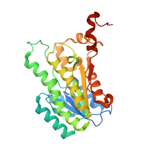 | |
UniProt | |||||
Find proteins for Q13GR0 (Paraburkholderia xenovorans (strain LB400)) Explore Q13GR0 Go to UniProtKB: Q13GR0 | |||||
Entity Groups | |||||
| Sequence Clusters | 30% Identity50% Identity70% Identity90% Identity95% Identity100% Identity | ||||
| UniProt Group | Q13GR0 | ||||
Sequence AnnotationsExpand | |||||
| |||||
| Ligands 1 Unique | |||||
|---|---|---|---|---|---|
| ID | Chains | Name / Formula / InChI Key | 2D Diagram | 3D Interactions | |
| NAD Query on NAD | E [auth A], F [auth B], G [auth C], H [auth D] | NICOTINAMIDE-ADENINE-DINUCLEOTIDE C21 H27 N7 O14 P2 BAWFJGJZGIEFAR-NNYOXOHSSA-N |  | ||
| Length ( Å ) | Angle ( ˚ ) |
|---|---|
| a = 126.48 | α = 90 |
| b = 109.96 | β = 93.9 |
| c = 64.99 | γ = 90 |
| Software Name | Purpose |
|---|---|
| SCALA | data scaling |
| PHENIX | refinement |
| PDB_EXTRACT | data extraction |
| XDS | data reduction |
| MoRDa | phasing |
RCSB PDB (citation) is hosted by
RCSB PDB is a member of the

