5J8Q

Crystal Structure of the Cysteine Desulfurase SufS of Bacillus subtilis


Experimental Data Snapshot

  • Method: X-RAY DIFFRACTION
  • Resolution: 1.70 Å
  • R-Value Free: 0.148 
  • R-Value Work: 0.116 
  • R-Value Observed: 0.118 

wwPDB Validation   3D Report Full Report


Ligand Structure Quality Assessment 


This is version 2.1 of the entry. See complete history


Literature

Crystal Structure of Bacillus subtilis Cysteine Desulfurase SufS and Its Dynamic Interaction with Frataxin and Scaffold Protein SufU.

Blauenburg, B.Mielcarek, A.Altegoer, F.Fage, C.D.Linne, U.Bange, G.Marahiel, M.A.

(2016) PLoS One 11: e0158749-e0158749

  • DOI: https://doi.org/10.1371/journal.pone.0158749
  • Primary Citation of Related Structures:  
    5J8Q

  • PubMed Abstract: 

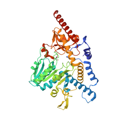
    The biosynthesis of iron sulfur (Fe-S) clusters in Bacillus subtilis is mediated by a SUF-type gene cluster, consisting of the cysteine desulfurase SufS, the scaffold protein SufU, and the putative chaperone complex SufB/SufC/SufD. Here, we present the high-resolution crystal structure of the SufS homodimer in its product-bound state (i.e., in complex with pyrodoxal-5'-phosphate, alanine, Cys361-persulfide). By performing hydrogen/deuterium exchange (H/DX) experiments, we characterized the interaction of SufS with SufU and demonstrate that SufU induces an opening of the active site pocket of SufS. Recent data indicate that frataxin could be involved in Fe-S cluster biosynthesis by facilitating iron incorporation. H/DX experiments show that frataxin indeed interacts with the SufS/SufU complex at the active site. Our findings deepen the current understanding of Fe-S cluster biosynthesis, a complex yet essential process, in the model organism B. subtilis.


  • Organizational Affiliation

    Department of Chemistry, Biochemistry, Hans-Meerwein Str. 4, Philipps University Marburg, 35043 Marburg, Germany.


Macromolecules
Find similar proteins by:  (by identity cutoff)  |  3D Structure
Entity ID: 1
MoleculeChains Sequence LengthOrganismDetailsImage
Cysteine desulfurase SufS414Bacillus subtilis subsp. subtilis str. 168Mutation(s): 0 
Gene Names: sufScsdyurWBSU32690
EC: 2.8.1.7
UniProt
Find proteins for O32164 (Bacillus subtilis (strain 168))
Explore O32164 
Go to UniProtKB:  O32164
Entity Groups  
Sequence Clusters30% Identity50% Identity70% Identity90% Identity95% Identity100% Identity
UniProt GroupO32164
Sequence Annotations
Expand
  • Reference Sequence
Small Molecules
Ligands 2 Unique
IDChains Name / Formula / InChI Key2D Diagram3D Interactions
PLP
Query on PLP

Download Ideal Coordinates CCD File 
B [auth A]PYRIDOXAL-5'-PHOSPHATE
C8 H10 N O6 P
NGVDGCNFYWLIFO-UHFFFAOYSA-N
ALA
Query on ALA

Download Ideal Coordinates CCD File 
C [auth A]ALANINE
C3 H7 N O2
QNAYBMKLOCPYGJ-REOHCLBHSA-N
Modified Residues  1 Unique
IDChains TypeFormula2D DiagramParent
CSS
Query on CSS
A
L-PEPTIDE LINKINGC3 H7 N O2 S2CYS
Experimental Data & Validation

Experimental Data

  • Method: X-RAY DIFFRACTION
  • Resolution: 1.70 Å
  • R-Value Free: 0.148 
  • R-Value Work: 0.116 
  • R-Value Observed: 0.118 
  • Space Group: P 31 2 1
Unit Cell:
Length ( Å )Angle ( ˚ )
a = 93.191α = 90
b = 93.191β = 90
c = 130.543γ = 120
Software Package:
Software NamePurpose
XDSdata reduction
Aimlessdata scaling
PHASERphasing
REFMACrefinement

Structure Validation

View Full Validation Report


Ligand Structure Quality Assessment 


Entry History 

Deposition Data

Revision History  (Full details and data files)

  • Version 1.0: 2016-07-27
    Type: Initial release
  • Version 2.0: 2020-01-15
    Changes: Atomic model
  • Version 2.1: 2024-01-10
    Changes: Data collection, Database references, Derived calculations, Refinement description