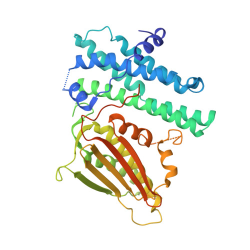
5J6A

Crystal structure of pyruvate dehydrogenase kinase isoform 2 in complex with inhibitor PS46


Experimental Data Snapshot

  • Method: X-RAY DIFFRACTION
  • Resolution: 2.04 Å
  • R-Value Free: 0.229 
  • R-Value Work: 0.196 
  • R-Value Observed: 0.198 

wwPDB Validation   3D Report Full Report


Ligand Structure Quality Assessment 


This is version 1.4 of the entry. See complete history


Literature

Development of Dihydroxyphenyl Sulfonylisoindoline Derivatives as Liver-Targeting Pyruvate Dehydrogenase Kinase Inhibitors.

Tso, S.C.Lou, M.Wu, C.Y.Gui, W.J.Chuang, J.L.Morlock, L.K.Williams, N.S.Wynn, R.M.Qi, X.Chuang, D.T.

(2017) J Med Chem 60: 1142-1150

  • DOI: https://doi.org/10.1021/acs.jmedchem.6b01540
  • Primary Citation of Related Structures:  
    5J6A, 5J71

  • PubMed Abstract: 

    Pyruvate dehydrogenase kinases 1-4 (PDK1-4) negatively control activity of the pyruvate dehydrogenase complex (PDC) and are up-regulated in obesity, diabetes, heart failure, and cancer. We reported earlier two novel pan-PDK inhibitors PS8 [4-((5-hydroxyisoindolin-2-yl)sulfonyl)benzene-1,3-diol] (1) and PS10 [2-((2,4-dihydroxyphenyl)sulfonyl)isoindoline-4,6-diol] (2) that targeted the ATP-binding pocket in PDKs. Here, we developed a new generation of PDK inhibitors by extending the dihydroxyphenyl sulfonylisoindoline scaffold in 1 and 2 to the entrance region of the ATP-binding pocket in PDK2. The lead inhibitor (S)-3-amino-4-(4-((2-((2,4-dihydroxyphenyl)sulfonyl)isoindolin-5-yl)amino)piperidin-1-yl)-4-oxobutanamide (17) shows a ∼8-fold lower IC 50 (58 nM) than 2 (456 nM). In the crystal structure, the asparagine moiety in 17 provides additional interactions with Glu-262 from PDK2. Treatment of diet-induced obese mice with 17 resulted in significant liver-specific augmentation of PDC activity, accompanied by improved glucose tolerance and drastically reduced hepatic steatosis. These findings support 17 as a potential glucose-lowering therapeutic targeting liver for obesity and type 2 diabetes.


  • Organizational Affiliation

    Chemistry Center, National Institute of Biological Science , Beijing 102206, China.


Macromolecules
Find similar proteins by:  (by identity cutoff)  |  3D Structure
Entity ID: 1
MoleculeChains Sequence LengthOrganismDetailsImage
[Pyruvate dehydrogenase (acetyl-transferring)] kinase isozyme 2, mitochondrial400Homo sapiensMutation(s): 0 
Gene Names: PDK2PDHK2
EC: 2.7.11.2
UniProt & NIH Common Fund Data Resources
Find proteins for Q15119 (Homo sapiens)
Explore Q15119 
Go to UniProtKB:  Q15119
PHAROS:  Q15119
GTEx:  ENSG00000005882 
Entity Groups  
Sequence Clusters30% Identity50% Identity70% Identity90% Identity95% Identity100% Identity
UniProt GroupQ15119
Sequence Annotations
Expand
  • Reference Sequence
Small Molecules
Ligands 1 Unique
IDChains Name / Formula / InChI Key2D Diagram3D Interactions
P46
Query on P46

Download Ideal Coordinates CCD File 
B [auth A](3S)-3-amino-4-[4-({2-[(2,4-dihydroxyphenyl)sulfonyl]-2H-isoindol-5-yl}amino)piperidin-1-yl]-4-oxobutanamide
C23 H27 N5 O6 S
HAMJZOUVHDEUSM-IBGZPJMESA-N
Binding Affinity Annotations 
IDSourceBinding Affinity
P46 Binding MOAD:  5J6A Kd: 22 (nM) from 1 assay(s)
Experimental Data & Validation

Experimental Data

  • Method: X-RAY DIFFRACTION
  • Resolution: 2.04 Å
  • R-Value Free: 0.229 
  • R-Value Work: 0.196 
  • R-Value Observed: 0.198 
  • Space Group: P 64
Unit Cell:
Length ( Å )Angle ( ˚ )
a = 109.389α = 90
b = 109.389β = 90
c = 84.42γ = 120
Software Package:
Software NamePurpose
PHENIXrefinement
HKL-3000data reduction
HKL-3000data scaling
PHENIXphasing

Structure Validation

View Full Validation Report


Ligand Structure Quality Assessment 


Entry History & Funding Information

Deposition Data


Funding OrganizationLocationGrant Number
National Institutes of Health/National Institute of Diabetes and Digestive and Kidney Disease (NIH/NIDDK)United StatesDK-62306
Welch FoundationUnited StatesI-1286

Revision History  (Full details and data files)

  • Version 1.0: 2017-01-25
    Type: Initial release
  • Version 1.1: 2017-02-22
    Changes: Database references
  • Version 1.2: 2017-09-20
    Changes: Author supporting evidence
  • Version 1.3: 2019-12-25
    Changes: Author supporting evidence
  • Version 1.4: 2023-09-27
    Changes: Data collection, Database references, Refinement description