5IEP

Structure of CDL2.3b, a computationally designed Vitamin-D3 binder


Experimental Data Snapshot

  • Method: X-RAY DIFFRACTION
  • Resolution: 1.89 Å
  • R-Value Free: 0.244 
  • R-Value Work: 0.228 
  • R-Value Observed: 0.230 

wwPDB Validation   3D Report Full Report


Ligand Structure Quality Assessment 


This is version 1.5 of the entry. See complete history


Literature

Unintended specificity of an engineered ligand-binding protein facilitated by unpredicted plasticity of the protein fold.

Day, A.L.Greisen, P.Doyle, L.Schena, A.Stella, N.Johnsson, K.Baker, D.Stoddard, B.

(2018) Protein Eng Des Sel 31: 375-387

  • DOI: https://doi.org/10.1093/protein/gzy031
  • Primary Citation of Related Structures:  
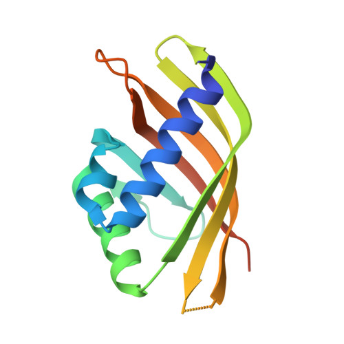
    5IEN, 5IEO, 5IEP

  • PubMed Abstract: 

    Attempts to create novel ligand-binding proteins often focus on formation of a binding pocket with shape complementarity against the desired ligand (particularly for compounds that lack distinct polar moieties). Although designed proteins often exhibit binding of the desired ligand, in some cases they display unintended recognition behavior. One such designed protein, that was originally intended to bind tetrahydrocannabinol (THC), was found instead to display binding of 25-hydroxy-cholecalciferol (25-D3) and was subjected to biochemical characterization, further selections for enhanced 25-D3 binding affinity and crystallographic analyses. The deviation in specificity is due in part to unexpected altertion of its conformation, corresponding to a significant change of the orientation of an α-helix and an equally large movement of a loop, both of which flank the designed ligand-binding pocket. Those changes led to engineered protein constructs that exhibit significantly more contacts and complementarity towards the 25-D3 ligand than the initial designed protein had been predicted to form towards its intended THC ligand. Molecular dynamics simulations imply that the initial computationally designed mutations may contribute to the movement of the helix. These analyses collectively indicate that accurate prediction and control of backbone dynamics conformation, through a combination of improved conformational sampling and/or de novo structure design, represents a key area of further development for the design and optimization of engineered ligand-binding proteins.


  • Organizational Affiliation

    Departments of Bioengineering and Biochemistry, University of Washington, Molecular Engineering and Sciences, Seattle, WA, USA.


Macromolecules
Find similar proteins by:  (by identity cutoff)  |  3D Structure
Entity ID: 1
MoleculeChains Sequence LengthOrganismDetailsImage
CDL2.3b137synthetic constructMutation(s): 0 
Entity Groups  
Sequence Clusters30% Identity50% Identity70% Identity90% Identity95% Identity100% Identity
Sequence Annotations
Expand
  • Reference Sequence
Small Molecules
Ligands 1 Unique
IDChains Name / Formula / InChI Key2D Diagram3D Interactions
VDY
Query on VDY

Download Ideal Coordinates CCD File 
B [auth A]3-{2-[1-(5-HYDROXY-1,5-DIMETHYL-HEXYL)-7A-METHYL-OCTAHYDRO-INDEN-4-YLIDENE]-ETHYLIDENE}-4-METHYLENE-CYCLOHEXANOL
C27 H44 O2
JWUBBDSIWDLEOM-DTOXIADCSA-N
Experimental Data & Validation

Experimental Data

  • Method: X-RAY DIFFRACTION
  • Resolution: 1.89 Å
  • R-Value Free: 0.244 
  • R-Value Work: 0.228 
  • R-Value Observed: 0.230 
  • Space Group: P 41 21 2
Unit Cell:
Length ( Å )Angle ( ˚ )
a = 71.848α = 90
b = 71.848β = 90
c = 60.787γ = 90
Software Package:
Software NamePurpose
PHENIXrefinement
HKL-2000data reduction
HKL-2000data scaling
PHASERphasing
PDB_EXTRACTdata extraction

Structure Validation

View Full Validation Report


Ligand Structure Quality Assessment 


Entry History & Funding Information

Deposition Data


Funding OrganizationLocationGrant Number
National Institutes of Health/National Institute of General Medical Sciences (NIH/NIGMS)United StatesGM115545

Revision History  (Full details and data files)

  • Version 1.0: 2017-03-01
    Type: Initial release
  • Version 1.1: 2017-09-27
    Changes: Author supporting evidence
  • Version 1.2: 2018-07-18
    Changes: Data collection, Database references, Structure summary
  • Version 1.3: 2019-12-25
    Changes: Author supporting evidence
  • Version 1.4: 2020-02-12
    Changes: Database references
  • Version 1.5: 2023-09-27
    Changes: Data collection, Database references, Refinement description