Recognition of the disordered p53 transactivation domain by the transcriptional adapter zinc finger domains of CREB-binding protein.
Krois, A.S., Ferreon, J.C., Martinez-Yamout, M.A., Dyson, H.J., Wright, P.E.(2016) Proc Natl Acad Sci U S A 113: E1853-E1862
- PubMed: 26976603
- DOI: https://doi.org/10.1073/pnas.1602487113
- Primary Citation of Related Structures:
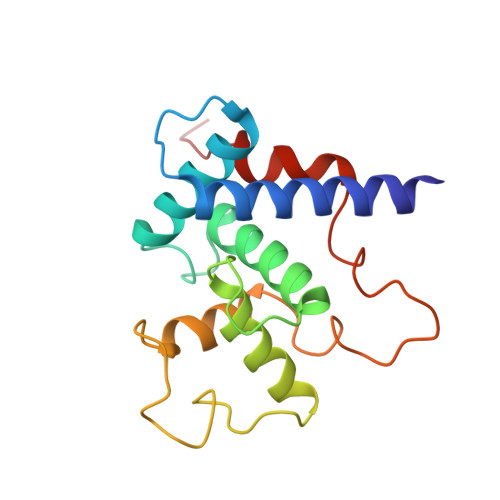
5HOU, 5HP0, 5HPD - PubMed Abstract:
An important component of the activity of p53 as a tumor suppressor is its interaction with the transcriptional coactivators cyclic-AMP response element-binding protein (CREB)-binding protein (CBP) and p300, which activate transcription of p53-regulated stress response genes and stabilize p53 against ubiquitin-mediated degradation. The highest affinity interactions are between the intrinsically disordered N-terminal transactivation domain (TAD) of p53 and the TAZ1 and TAZ2 domains of CBP/p300. The NMR spectra of simple binary complexes of the TAZ1 and TAZ2 domains with the p53TAD suffer from exchange broadening, but innovations in construct design and isotopic labeling have enabled us to obtain high-resolution structures using fusion proteins, uniformly labeled in the case of the TAZ2-p53TAD fusion and segmentally labeled through transintein splicing for the TAZ1-p53TAD fusion. The p53TAD is bipartite, with two interaction motifs, termed AD1 and AD2, which fold to form short amphipathic helices upon binding to TAZ1 and TAZ2 whereas intervening regions of the p53TAD remain flexible. Both the AD1 and AD2 motifs bind to hydrophobic surfaces of the TAZ domains, with AD2 making more extensive hydrophobic contacts consistent with its greater contribution to the binding affinity. Binding of AD1 and AD2 is synergistic, and structural studies performed with isolated motifs can be misleading. The present structures of the full-length p53TAD complexes demonstrate the versatility of the interactions available to an intrinsically disordered domain containing bipartite interaction motifs and provide valuable insights into the structural basis of the affinity changes that occur upon stress-related posttranslational modification.
Organizational Affiliation:
Department of Integrative Structural and Computational Biology, The Scripps Research Institute, La Jolla, CA 92037; Skaggs Institute for Chemical Biology, The Scripps Research Institute, La Jolla, CA 92037.