5H8S

Structure of the human GluA2 LBD in complex with GNE3419


Experimental Data Snapshot

  • Method: X-RAY DIFFRACTION
  • Resolution: 1.70 Å
  • R-Value Free: 0.206 
  • R-Value Work: 0.174 
  • R-Value Observed: 0.176 

wwPDB Validation   3D Report Full Report


Ligand Structure Quality Assessment 


This is version 1.2 of the entry. See complete history


Literature

Positive Allosteric Modulators of GluN2A-Containing NMDARs with Distinct Modes of Action and Impacts on Circuit Function.

Hackos, D.H.Lupardus, P.J.Grand, T.Chen, Y.Wang, T.M.Reynen, P.Gustafson, A.Wallweber, H.J.Volgraf, M.Sellers, B.D.Schwarz, J.B.Paoletti, P.Sheng, M.Zhou, Q.Hanson, J.E.

(2016) Neuron 89: 983-999

  • DOI: https://doi.org/10.1016/j.neuron.2016.01.016
  • Primary Citation of Related Structures:  
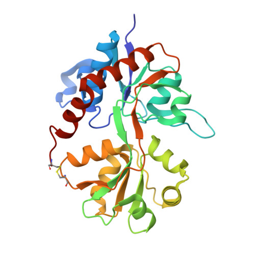
    5H8F, 5H8H, 5H8N, 5H8Q, 5H8S, 5KCJ

  • PubMed Abstract: 

    To enhance physiological function of NMDA receptors (NMDARs), we identified positive allosteric modulators (PAMs) of NMDARs with selectivity for GluN2A subunit-containing receptors. X-ray crystallography revealed a binding site at the GluN1-GluN2A dimer interface of the extracellular ligand-binding domains (LBDs). Despite the similarity between the LBDs of NMDARs and AMPA receptors (AMPARs), GluN2A PAMs with good selectivity against AMPARs were identified. Potentiation was observed with recombinant triheteromeric GluN1/GluN2A/GluN2B NMDARs and with synaptically activated NMDARs in brain slices from wild-type (WT), but not GluN2A knockout (KO), mice. Individual GluN2A PAMs exhibited variable degrees of glutamate (Glu) dependence, impact on NMDAR Glu EC50, and slowing of channel deactivation. These distinct PAMs also exhibited differential impacts during synaptic plasticity induction. The identification of a new NMDAR modulatory site and characterization of GluN2A-selective PAMs provide powerful molecular tools to dissect NMDAR function and demonstrate the feasibility of a therapeutically desirable type of NMDAR enhancement.


  • Organizational Affiliation

    Department of Neuroscience, Genentech, Inc., 1 DNA Way, South San Francisco, CA 94080, USA.


Macromolecules
Find similar proteins by:  (by identity cutoff)  |  3D Structure
Entity ID: 1
MoleculeChains Sequence LengthOrganismDetailsImage
Glutamate receptor 2,Glutamate receptor 2A,
B [auth C],
C [auth B]
263Homo sapiensMutation(s): 2 
Gene Names: GRIA2GLUR2
UniProt & NIH Common Fund Data Resources
Find proteins for P42262 (Homo sapiens)
Explore P42262 
Go to UniProtKB:  P42262
PHAROS:  P42262
GTEx:  ENSG00000120251 
Entity Groups  
Sequence Clusters30% Identity50% Identity70% Identity90% Identity95% Identity100% Identity
UniProt GroupP42262
Sequence Annotations
Expand
  • Reference Sequence
Small Molecules
Ligands 4 Unique
IDChains Name / Formula / InChI Key2D Diagram3D Interactions
5YC
Query on 5YC

Download Ideal Coordinates CCD File 
E [auth A],
J [auth C]
7-[[ethyl(phenyl)amino]methyl]-2-methyl-[1,3,4]thiadiazolo[3,2-a]pyrimidin-5-one
C15 H16 N4 O S
MAVHHQOKQQPPKJ-UHFFFAOYSA-N
GLU
Query on GLU

Download Ideal Coordinates CCD File 
D [auth A],
I [auth C],
M [auth B]
GLUTAMIC ACID
C5 H9 N O4
WHUUTDBJXJRKMK-VKHMYHEASA-N
CAC
Query on CAC

Download Ideal Coordinates CCD File 
H [auth A]CACODYLATE ION
C2 H6 As O2
OGGXGZAMXPVRFZ-UHFFFAOYSA-M
ZN
Query on ZN

Download Ideal Coordinates CCD File 
F [auth A]
G [auth A]
K [auth C]
L [auth C]
N [auth B]
F [auth A],
G [auth A],
K [auth C],
L [auth C],
N [auth B],
O [auth B]
ZINC ION
Zn
PTFCDOFLOPIGGS-UHFFFAOYSA-N
Binding Affinity Annotations 
IDSourceBinding Affinity
5YC BindingDB:  5H8S EC50: min: 2000, max: 7500 (nM) from 2 assay(s)
Experimental Data & Validation

Experimental Data

  • Method: X-RAY DIFFRACTION
  • Resolution: 1.70 Å
  • R-Value Free: 0.206 
  • R-Value Work: 0.174 
  • R-Value Observed: 0.176 
  • Space Group: P 2 21 21
Unit Cell:
Length ( Å )Angle ( ˚ )
a = 47.232α = 90
b = 114.461β = 90
c = 162.744γ = 90
Software Package:
Software NamePurpose
PHENIXrefinement
HKL-2000data reduction
SCALEPACKdata scaling
PHASERphasing

Structure Validation

View Full Validation Report


Ligand Structure Quality Assessment 


Entry History 

Deposition Data

Revision History  (Full details and data files)

  • Version 1.0: 2016-02-24
    Type: Initial release
  • Version 1.1: 2018-04-18
    Changes: Data collection, Database references, Derived calculations
  • Version 1.2: 2023-09-27
    Changes: Data collection, Database references, Derived calculations, Refinement description