5GX9

PYP mutant - E46Q


Experimental Data Snapshot

  • Method: NEUTRON DIFFRACTION
  • Resolution: 1.49 Å
  • R-Value Free: 0.200 
  • R-Value Work: 0.158 
  • R-Value Observed: 0.162 

wwPDB Validation   3D Report Full Report


This is version 1.3 of the entry. See complete history


Literature

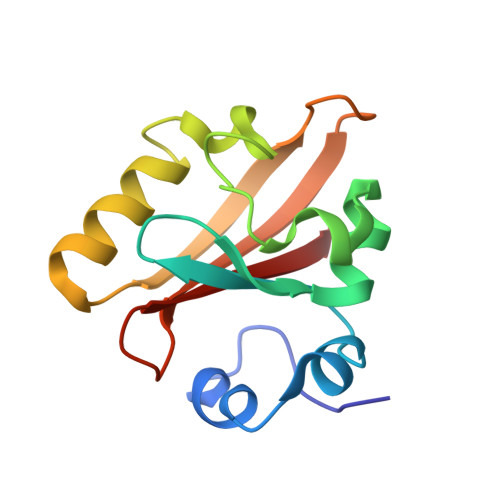
Neutron crystallography of photoactive yellow protein reveals unusual protonation state of Arg52 in the crystal

Yonezawa, K.Shimizu, N.Kurihara, K.Yamazaki, Y.Kamikubo, H.Kataoka, M.

(2017) Sci Rep 7: 9361-9361

  • DOI: https://doi.org/10.1038/s41598-017-09718-9
  • Primary Citation of Related Structures:  
    5GX9

  • PubMed Abstract: 

    Because of its high pK a , arginine (Arg) is believed to be protonated even in the hydrophobic environment of the protein interior. However, our neutron crystallographic structure of photoactive yellow protein, a light sensor, demonstrated that Arg52 adopts an electrically neutral form. We also showed that the hydrogen bond between the chromophore and Glu46 is a so-called low barrier hydrogen bond (LBHB). Because both the neutral Arg and LBHB are unusual in proteins, these observations remain controversial. To validate our findings, we carried out neutron crystallographic analysis of the E46Q mutant of PYP. The resultant structure revealed that the proportion of the cationic form is higher in E46Q than in WT, although the cationic and neutral forms of Arg52 coexist in E46Q. These observations were confirmed by the occupancy of the deuterium atom bound to the N η1 atom combined with an alternative conformation of the N (η2) D 2 group comprising sp 2 hybridisation. Based on these results, we propose that the formation of the LBHB decreases the proton affinity of Arg52, stabilizing the neutral form in the crystal.


  • Organizational Affiliation

    Graduate School of Materials Science, Nara Institute of Science and Technology, 8916-5 Takayama, Ikoma, Nara, 630-0192, Japan.


Macromolecules
Find similar proteins by:  (by identity cutoff)  |  3D Structure
Entity ID: 1
MoleculeChains Sequence LengthOrganismDetailsImage
Photoactive yellow protein125Halorhodospira halophilaMutation(s): 1 
Gene Names: pyp
UniProt
Find proteins for P16113 (Halorhodospira halophila)
Explore P16113 
Go to UniProtKB:  P16113
Entity Groups  
Sequence Clusters30% Identity50% Identity70% Identity90% Identity95% Identity100% Identity
UniProt GroupP16113
Sequence Annotations
Expand
  • Reference Sequence
Small Molecules
Ligands 1 Unique
IDChains Name / Formula / InChI Key2D Diagram3D Interactions
HC4
Query on HC4

Download Ideal Coordinates CCD File 
B [auth A]4'-HYDROXYCINNAMIC ACID
C9 H8 O3
NGSWKAQJJWESNS-ZZXKWVIFSA-N
Experimental Data & Validation

Experimental Data

  • Method: NEUTRON DIFFRACTION
  • Resolution: 1.49 Å
  • R-Value Free: 0.200 
  • R-Value Work: 0.158 
  • R-Value Observed: 0.162 
  • Space Group: P 63
Unit Cell:
Length ( Å )Angle ( ˚ )
a = 66.823α = 90
b = 66.823β = 90
c = 40.982γ = 120
Software Package:
Software NamePurpose
PHENIXrefinement
AMoREmodel building

Structure Validation

View Full Validation Report


Entry History & Funding Information

Deposition Data

  • Released Date: 2017-08-30 
  • Deposition Author(s): Yonezawa, K.

Funding OrganizationLocationGrant Number
Japanese Ministry of Education, Culture, Sports, Science and TechnologyJapan25102003

Revision History  (Full details and data files)

  • Version 1.0: 2017-08-30
    Type: Initial release
  • Version 1.1: 2017-09-13
    Changes: Database references
  • Version 1.2: 2018-11-28
    Changes: Data collection
  • Version 1.3: 2023-11-08
    Changes: Data collection, Database references, Refinement description