5GK1

Crystal structure of the ketosynthase StlD complexed with substrate


Experimental Data Snapshot

  • Method: X-RAY DIFFRACTION
  • Resolution: 1.83 Å
  • R-Value Free: 0.231 
  • R-Value Work: 0.198 
  • R-Value Observed: 0.200 

wwPDB Validation   3D Report Full Report


This is version 1.2 of the entry. See complete history


Literature

Structural Insight into the Enzymatic Formation of Bacterial Stilbene.

Mori, T.Awakawa, T.Shimomura, K.Saito, Y.Yang, D.Morita, H.Abe, I.

(2016) Cell Chem Biol 23: 1468-1479

  • DOI: https://doi.org/10.1016/j.chembiol.2016.10.010
  • Primary Citation of Related Structures:  
    5GK0, 5GK1, 5GK2

  • PubMed Abstract: 

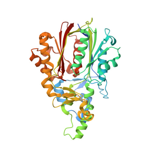
    In contrast to stilbene biosynthesis by type III polyketide synthase in plants, in bacteria stilbene is produced by the collaboration of two enzymes in Photorhabdus luminescens: the unusual β-ketosynthase StlD catalyzes the condensation of the β-ketoacyl starter with an α,β-unsaturated-acyl substrate (two C-C bond-forming reactions) to produce isopropylstyrylcyclohexanedione, which is subsequently converted to stilbene by the aromatase StlC. Here we report the in vitro characterizations of StlD and StlC, and the X-ray crystal structures of StlD. Interestingly, structure-based mutagenesis demonstrated that His302, within the conserved Cys-His-Asn triad, is not essential for the enzyme reaction, while Glu154 functions as a base-catalyst to activate the β-ketoacyl intermediate bound to the catalytic Cys126. The structures also revealed the presence of a putative nucleophilic water molecule activated by hydrogen bond networks with Glu154 and Ser340, suggesting that StlD employs novel catalytic machinery for the condensation of two acyl substrates to produce the cyclohexanedione scaffold.


  • Organizational Affiliation

    Graduate School of Pharmaceutical Sciences, The University of Tokyo, 7-3-1 Hongo, Bunkyo-ku, Tokyo 113-0033, Japan.


Macromolecules
Find similar proteins by:  (by identity cutoff)  |  3D Structure
Entity ID: 1
MoleculeChains Sequence LengthOrganismDetailsImage
Ketosynthase StlD
A, B, C, D
402Photorhabdus laumondii subsp. laumondii TTO1Mutation(s): 0 
Gene Names: plu2164
UniProt
Find proteins for Q7N4Z6 (Photorhabdus laumondii subsp. laumondii (strain DSM 15139 / CIP 105565 / TT01))
Explore Q7N4Z6 
Go to UniProtKB:  Q7N4Z6
Entity Groups  
Sequence Clusters30% Identity50% Identity70% Identity90% Identity95% Identity100% Identity
UniProt GroupQ7N4Z6
Sequence Annotations
Expand
  • Reference Sequence
Small Molecules
Experimental Data & Validation

Experimental Data

  • Method: X-RAY DIFFRACTION
  • Resolution: 1.83 Å
  • R-Value Free: 0.231 
  • R-Value Work: 0.198 
  • R-Value Observed: 0.200 
  • Space Group: P 1 21 1
Unit Cell:
Length ( Å )Angle ( ˚ )
a = 63.29α = 90
b = 127.64β = 91.55
c = 107.89γ = 90
Software Package:
Software NamePurpose
PHENIXrefinement
XDSdata reduction
XDSdata scaling
PHASERphasing

Structure Validation

View Full Validation Report


Entry History 

Deposition Data

Revision History  (Full details and data files)

  • Version 1.0: 2017-07-05
    Type: Initial release
  • Version 1.1: 2019-01-16
    Changes: Data collection, Database references
  • Version 1.2: 2023-11-08
    Changes: Data collection, Database references, Refinement description