5G5R

CBS domain tandem of site-2 protease from Archaeoglobus fulgidus in complex with llama Nanobody - apo form


Experimental Data Snapshot

  • Method: X-RAY DIFFRACTION
  • Resolution: 2.40 Å
  • R-Value Free: 0.250 
  • R-Value Work: 0.207 
  • R-Value Observed: 0.209 

wwPDB Validation   3D Report Full Report


This is version 1.2 of the entry. See complete history


Literature

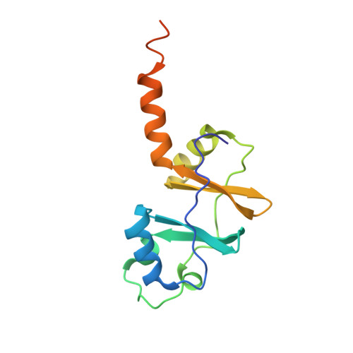
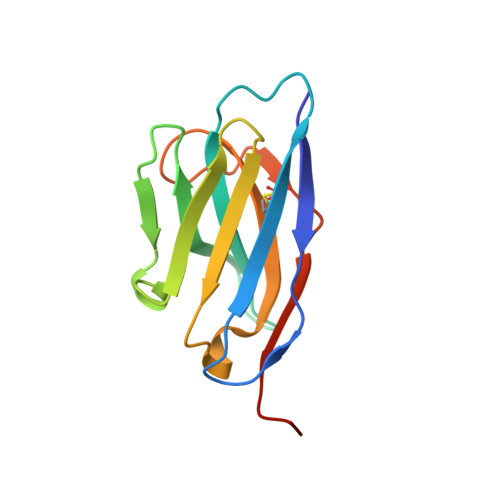
Crystallographic and biochemical characterization of the dimeric architecture of site-2 protease.

Schacherl, M.Gompert, M.Pardon, E.Lamkemeyer, T.Steyaert, J.Baumann, U.

(2017) Biochim Biophys Acta 1859: 1859-1871

  • DOI: https://doi.org/10.1016/j.bbamem.2017.05.006
  • Primary Citation of Related Structures:  
    5G5R, 5G5X

  • PubMed Abstract: 

    Regulated intramembrane proteolysis by members of the site-2 protease family (S2P) is an essential signal transduction mechanism conserved from bacteria to humans. There is some evidence that extra-membranous domains, like PDZ and CBS domains, regulate the proteolytic activity of S2Ps and that some members act as dimers. Here we report the crystal structure of the regulatory CBS domain pair of S2P from Archaeoglobus fulgidus, AfS2P, in the apo and nucleotide-bound form in complex with a specific nanobody from llama. Cross-linking and SEC-MALS analyses show for the first time the dimeric architecture of AfS2P both in the membrane and in detergent micelles. The CBS domain pair dimer (CBS module) displays an unusual head-to-tail configuration and nucleotide binding triggers no major conformational changes in the magnesium-free state. In solution, MgATP drives monomerization of the CBS module. We propose a model of the so far unknown architecture of the transmembrane domain dimer and for a regulatory mechanism of AfS2P that involves the interaction of positively charged arginine residues located at the cytoplasmic face of the transmembrane domain with the negatively charged phosphate groups of ATP moieties bound to the CBS domain pairs. Binding of MgATP could promote opening of the CBS module to allow lateral access of the globular cytoplasmic part of the substrate.


  • Organizational Affiliation

    Institute of Biochemistry, University of Cologne, Otto-Fischer-Str. 14, 50674 Cologne, Germany. Electronic address: magdalena.schacherl@uni-koeln.de.


Macromolecules
Find similar proteins by:  (by identity cutoff)  |  3D Structure
Entity ID: 1
MoleculeChains Sequence LengthOrganismDetailsImage
SITE-2 PROTEASE138Archaeoglobus fulgidusMutation(s): 0 
EC: 3.4.24.85
UniProt
Find proteins for O29915 (Archaeoglobus fulgidus (strain ATCC 49558 / DSM 4304 / JCM 9628 / NBRC 100126 / VC-16))
Explore O29915 
Go to UniProtKB:  O29915
Entity Groups  
Sequence Clusters30% Identity50% Identity70% Identity90% Identity95% Identity100% Identity
UniProt GroupO29915
Sequence Annotations
Expand
  • Reference Sequence
Find similar proteins by:  (by identity cutoff)  |  3D Structure
Entity ID: 2
MoleculeChains Sequence LengthOrganismDetailsImage
NANOBODY123Lama glamaMutation(s): 0 
Entity Groups  
Sequence Clusters30% Identity50% Identity70% Identity90% Identity95% Identity100% Identity
Sequence Annotations
Expand
  • Reference Sequence
Experimental Data & Validation

Experimental Data

  • Method: X-RAY DIFFRACTION
  • Resolution: 2.40 Å
  • R-Value Free: 0.250 
  • R-Value Work: 0.207 
  • R-Value Observed: 0.209 
  • Space Group: P 61 2 2
Unit Cell:
Length ( Å )Angle ( ˚ )
a = 68.44α = 90
b = 68.44β = 90
c = 196.8γ = 120
Software Package:
Software NamePurpose
PHENIXrefinement
XDSdata reduction
XSCALEdata scaling
PHASERphasing

Structure Validation

View Full Validation Report


Entry History 

Deposition Data

Revision History  (Full details and data files)

  • Version 1.0: 2017-05-24
    Type: Initial release
  • Version 1.1: 2017-07-19
    Changes: Database references
  • Version 1.2: 2024-01-10
    Changes: Data collection, Database references, Derived calculations, Refinement description