5FI5

HETEROYOHIMBINE SYNTHASE THAS1 FROM CATHARANTHUS ROSEUS - APO FORM


Experimental Data Snapshot

  • Method: X-RAY DIFFRACTION
  • Resolution: 2.25 Å
  • R-Value Free: 0.245 
  • R-Value Work: 0.203 
  • R-Value Observed: 0.205 

wwPDB Validation   3D Report Full Report


This is version 1.2 of the entry. See complete history


Literature

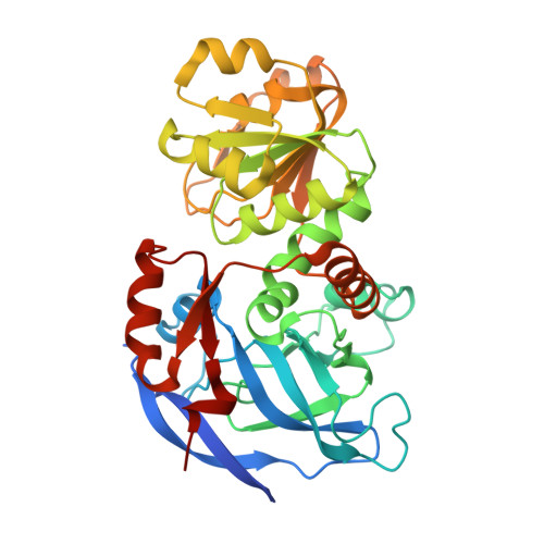
Structural investigation of heteroyohimbine alkaloid synthesis reveals active site elements that control stereoselectivity.

Stavrinides, A.Tatsis, E.C.Caputi, L.Foureau, E.Stevenson, C.E.Lawson, D.M.Courdavault, V.O'Connor, S.E.

(2016) Nat Commun 7: 12116-12116

  • DOI: https://doi.org/10.1038/ncomms12116
  • Primary Citation of Related Structures:  
    5FI3, 5FI5, 5H81, 5H82, 5H83

  • PubMed Abstract: 

    Plants produce an enormous array of biologically active metabolites, often with stereochemical variations on the same molecular scaffold. These changes in stereochemistry dramatically impact biological activity. Notably, the stereoisomers of the heteroyohimbine alkaloids show diverse pharmacological activities. We reported a medium chain dehydrogenase/reductase (MDR) from Catharanthus roseus that catalyses formation of a heteroyohimbine isomer. Here we report the discovery of additional heteroyohimbine synthases (HYSs), one of which produces a mixture of diastereomers. The crystal structures for three HYSs have been solved, providing insight into the mechanism of reactivity and stereoselectivity, with mutation of one loop transforming product specificity. Localization and gene silencing experiments provide a basis for understanding the function of these enzymes in vivo. This work sets the stage to explore how MDRs evolved to generate structural and biological diversity in specialized plant metabolism and opens the possibility for metabolic engineering of new compounds based on this scaffold.


  • Organizational Affiliation

    The John Innes Centre, Department of Biological Chemistry, Norwich NR4 7UH, UK.


Macromolecules
Find similar proteins by:  (by identity cutoff)  |  3D Structure
Entity ID: 1
MoleculeChains Sequence LengthOrganismDetailsImage
Tetrahydroalstonine synthase
A, B
357Catharanthus roseusMutation(s): 0 
UniProt
Find proteins for A0A0F6SD02 (Catharanthus roseus)
Explore A0A0F6SD02 
Go to UniProtKB:  A0A0F6SD02
Entity Groups  
Sequence Clusters30% Identity50% Identity70% Identity90% Identity95% Identity100% Identity
UniProt GroupA0A0F6SD02
Sequence Annotations
Expand
  • Reference Sequence
Experimental Data & Validation

Experimental Data

  • Method: X-RAY DIFFRACTION
  • Resolution: 2.25 Å
  • R-Value Free: 0.245 
  • R-Value Work: 0.203 
  • R-Value Observed: 0.205 
  • Space Group: P 31 2 1
Unit Cell:
Length ( Å )Angle ( ˚ )
a = 88.93α = 90
b = 88.93β = 90
c = 188.12γ = 120
Software Package:
Software NamePurpose
Aimlessdata scaling
REFMACrefinement
PDB_EXTRACTdata extraction
XDSdata reduction
PHASERphasing

Structure Validation

View Full Validation Report


Entry History & Funding Information

Deposition Data


Funding OrganizationLocationGrant Number
European Research CouncilUnited Kingdom311363
Biotechnology and Biological Sciences Research CouncilUnited KingdomBB/J004561/1
Region CentreFranceABISAL
Biotechnology and Biological Sciences Research CouncilUnited KingdomDoctoral Training Partnership

Revision History  (Full details and data files)

  • Version 1.0: 2016-07-27
    Type: Initial release
  • Version 1.1: 2017-08-30
    Changes: Author supporting evidence
  • Version 1.2: 2024-01-10
    Changes: Data collection, Database references, Refinement description