5F3X

Crystal structure of Harmonin NPDZ1 in complex with ANKS4B SAM-PBM


Experimental Data Snapshot

  • Method: X-RAY DIFFRACTION
  • Resolution: 2.65 Å
  • R-Value Free: 0.264 
  • R-Value Work: 0.227 
  • R-Value Observed: 0.229 

wwPDB Validation   3D Report Full Report


This is version 1.1 of the entry. See complete history


Literature

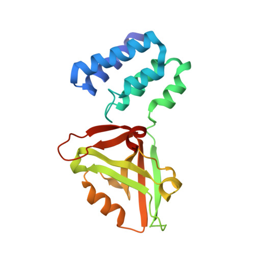
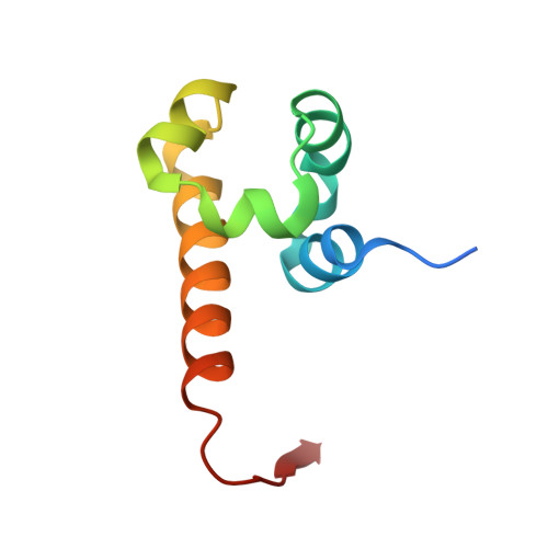
Mechanistic Basis of Organization of the Harmonin/USH1C-Mediated Brush Border Microvilli Tip-Link Complex

Li, J.He, Y.Lu, Q.Zhang, M.

(2016) Dev Cell 36: 179-189

  • DOI: https://doi.org/10.1016/j.devcel.2015.12.020
  • Primary Citation of Related Structures:  
    5F3X, 5F3Y

  • PubMed Abstract: 

    Brush border microvilli are actin-based protrusions lining the apical surface of epithelial cells in intestines and proximal tubules of kidneys. While brush border microvilli resemble the relatively well-characterized stereocilia of hair cells, the mechanistic basis of tip-link complex organization in microvilli is poorly understood. Here, we have biochemically and structurally characterized the following pairs of interactions: protocadherin 24 and Harmonin (also known as USH1C or AIE-75), Harmonin and myosin VIIb (MYO7B), Harmonin and ANKS4B, and ANKS4B and MYO7B. We show that Harmonin, ANKS4B, and MYO7B form a stable ternary complex for anchoring microvilli tip-link cadherins. Despite having only Harmonin in common, the microvilli and the stereocilia tip-link complexes are formed via strikingly similar interaction modes. These results not only provide insight into the mechanistic bases of brush border microvilli formation and maintenance but may also be valuable for understanding some gut and/or kidney diseases caused by perturbations of brush border microvilli structures.


  • Organizational Affiliation

    Division of Life Science, State Key Laboratory of Molecular Neuroscience, Hong Kong University of Science and Technology, Clear Water Bay, Kowloon, Hong Kong, China.


Macromolecules
Find similar proteins by:  (by identity cutoff)  |  3D Structure
Entity ID: 1
MoleculeChains Sequence LengthOrganismDetailsImage
Harmonin
A, C
196Homo sapiensMutation(s): 0 
Gene Names: USH1CAIE75
UniProt & NIH Common Fund Data Resources
Find proteins for Q9Y6N9 (Homo sapiens)
Explore Q9Y6N9 
Go to UniProtKB:  Q9Y6N9
PHAROS:  Q9Y6N9
GTEx:  ENSG00000006611 
Entity Groups  
Sequence Clusters30% Identity50% Identity70% Identity90% Identity95% Identity100% Identity
UniProt GroupQ9Y6N9
Sequence Annotations
Expand
  • Reference Sequence
Find similar proteins by:  (by identity cutoff)  |  3D Structure
Entity ID: 2
MoleculeChains Sequence LengthOrganismDetailsImage
Ankyrin repeat and SAM domain-containing protein 4B
B, D
81Mus musculusMutation(s): 0 
Gene Names: Anks4bHarp
UniProt & NIH Common Fund Data Resources
Find proteins for Q8K3X6 (Mus musculus)
Explore Q8K3X6 
Go to UniProtKB:  Q8K3X6
IMPC:  MGI:1919324
Entity Groups  
Sequence Clusters30% Identity50% Identity70% Identity90% Identity95% Identity100% Identity
UniProt GroupQ8K3X6
Sequence Annotations
Expand
  • Reference Sequence
Experimental Data & Validation

Experimental Data

  • Method: X-RAY DIFFRACTION
  • Resolution: 2.65 Å
  • R-Value Free: 0.264 
  • R-Value Work: 0.227 
  • R-Value Observed: 0.229 
  • Space Group: P 32
Unit Cell:
Length ( Å )Angle ( ˚ )
a = 124.566α = 90
b = 124.566β = 90
c = 49.654γ = 120
Software Package:
Software NamePurpose
PHENIXrefinement
HKL-2000data reduction
HKL-2000data scaling
PHASERphasing

Structure Validation

View Full Validation Report


Entry History 

Deposition Data

Revision History  (Full details and data files)

  • Version 1.0: 2016-03-16
    Type: Initial release
  • Version 1.1: 2023-11-08
    Changes: Data collection, Database references, Derived calculations, Refinement description