X-ray Crystallographic Structure of Oligomers Formed by a Toxic beta-Hairpin Derived from alpha-Synuclein: Trimers and Higher-Order Oligomers.
Salveson, P.J., Spencer, R.K., Nowick, J.S.(2016) J Am Chem Soc 138: 4458-4467
- PubMed: 26926877
- DOI: https://doi.org/10.1021/jacs.5b13261
- Primary Citation of Related Structures:
5F1T, 5F1W - PubMed Abstract:
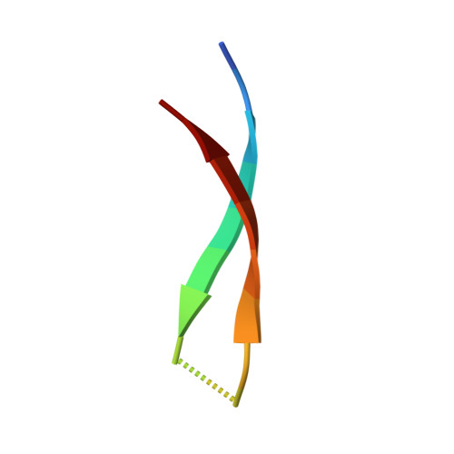
Oligomeric assemblies of the protein α-synuclein are thought to cause neurodegeneration in Parkinson's disease and related synucleinopathies. Characterization of α-synuclein oligomers at high resolution is an outstanding challenge in the field of structural biology. The absence of high-resolution structures of oligomers formed by α-synuclein impedes understanding the synucleinopathies at the molecular level. This paper reports the X-ray crystallographic structure of oligomers formed by a peptide derived from residues 36-55 of α-synuclein. The peptide 1a adopts a β-hairpin structure, which assembles in a hierarchical fashion. Three β-hairpins assemble to form a triangular trimer. Three copies of the triangular trimer assemble to form a basket-shaped nonamer. Two nonamers pack to form an octadecamer. Molecular modeling suggests that full-length α-synuclein may also be able to assemble in this fashion. Circular dichroism spectroscopy demonstrates that peptide 1a interacts with anionic lipid bilayer membranes, like oligomers of full-length α-synuclein. LDH and MTT assays demonstrate that peptide 1a is toxic toward SH-SY5Y cells. Comparison of peptide 1a to homologues suggests that this toxicity results from nonspecific interactions with the cell membrane. The oligomers formed by peptide 1a are fundamentally different than the proposed models of the fibrils formed by α-synuclein and suggest that α-Syn36-55, rather than the NAC, may nucleate oligomer formation.
Organizational Affiliation:
Department of Chemistry, University of California Irvine , Irvine, California 92697-2025, United States.