5EYH

Crystal Structure of IMPase/NADP phosphatase complexed with NADP and Ca2+ at pH 7.0


Experimental Data Snapshot

  • Method: X-RAY DIFFRACTION
  • Resolution: 2.50 Å
  • R-Value Free: 0.241 
  • R-Value Work: 0.181 
  • R-Value Observed: 0.184 

wwPDB Validation   3D Report Full Report


Ligand Structure Quality Assessment 


This is version 1.2 of the entry. See complete history


Literature

Structural elucidation of the NADP(H) phosphatase activity of staphylococcal dual-specific IMPase/NADP(H) phosphatase

Bhattacharyya, S.Dutta, A.Dutta, D.Ghosh, A.K.Das, A.K.

(2016) Acta Crystallogr D Struct Biol 72: 281-290

  • DOI: https://doi.org/10.1107/S2059798316000620
  • Primary Citation of Related Structures:  
    5EYG, 5EYH, 5F24

  • PubMed Abstract: 

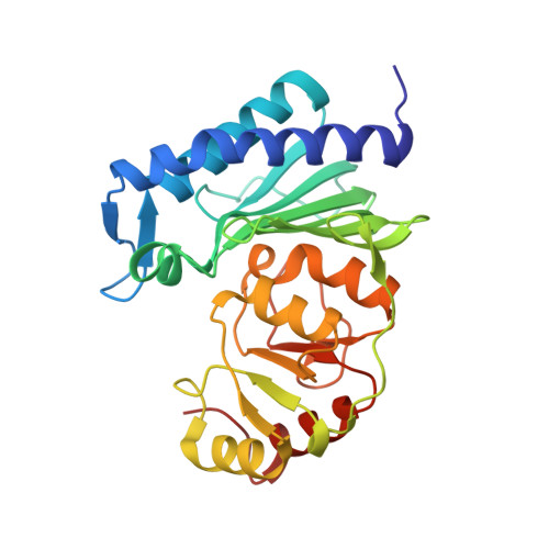
    NADP(H)/NAD(H) homeostasis has long been identified to play a pivotal role in the mitigation of reactive oxygen stress (ROS) in the intracellular milieu and is therefore critical for the progression and pathogenesis of many diseases. NAD(H) kinases and NADP(H) phosphatases are two key players in this pathway. Despite structural evidence demonstrating the existence and mode of action of NAD(H) kinases, the specific annotation and the mode of action of NADP(H) phosphatases remains obscure. Here, structural evidence supporting the alternative role of inositol monophosphatase (IMPase) as an NADP(H) phosphatase is reported. Crystal structures of staphylococcal dual-specific IMPase/NADP(H) phosphatase (SaIMPase-I) in complex with the substrates D-myo-inositol-1-phosphate and NADP(+) have been solved. The structure of the SaIMPase-I-Ca(2+)-NADP(+) ternary complex reveals the catalytic mode of action of NADP(H) phosphatase. Moreover, structures of SaIMPase-I-Ca(2+)-substrate complexes have reinforced the earlier proposal that the length of the active-site-distant helix α4 and its preceding loop are the predisposing factors for the promiscuous substrate specificity of SaIMPase-I. Altogether, the evidence presented suggests that IMPase-family enzymes with a shorter α4 helix could be potential candidates for previously unreported NADP(H) phosphatase activity.


  • Organizational Affiliation

    Department of Biotechnology, Indian Institute of Technology Kharagpur, Kharagpur 721 302, India.


Macromolecules
Find similar proteins by:  (by identity cutoff)  |  3D Structure
Entity ID: 1
MoleculeChains Sequence LengthOrganismDetailsImage
Inositol monophosphatase
A, B
265Staphylococcus aureusMutation(s): 1 
EC: 3.1.3.25
UniProt
Find proteins for Q2FVV7 (Staphylococcus aureus (strain NCTC 8325 / PS 47))
Explore Q2FVV7 
Go to UniProtKB:  Q2FVV7
Entity Groups  
Sequence Clusters30% Identity50% Identity70% Identity90% Identity95% Identity100% Identity
UniProt GroupQ2FVV7
Sequence Annotations
Expand
  • Reference Sequence
Small Molecules
Ligands 3 Unique
IDChains Name / Formula / InChI Key2D Diagram3D Interactions
NAP
Query on NAP

Download Ideal Coordinates CCD File 
H [auth A],
K [auth B]
NADP NICOTINAMIDE-ADENINE-DINUCLEOTIDE PHOSPHATE
C21 H28 N7 O17 P3
XJLXINKUBYWONI-NNYOXOHSSA-N
GOL
Query on GOL

Download Ideal Coordinates CCD File 
E [auth A],
F [auth A]
GLYCEROL
C3 H8 O3
PEDCQBHIVMGVHV-UHFFFAOYSA-N
CA
Query on CA

Download Ideal Coordinates CCD File 
C [auth A],
D [auth A],
G [auth A],
I [auth B],
J [auth B]
CALCIUM ION
Ca
BHPQYMZQTOCNFJ-UHFFFAOYSA-N
Experimental Data & Validation

Experimental Data

  • Method: X-RAY DIFFRACTION
  • Resolution: 2.50 Å
  • R-Value Free: 0.241 
  • R-Value Work: 0.181 
  • R-Value Observed: 0.184 
  • Space Group: P 1 21 1
Unit Cell:
Length ( Å )Angle ( ˚ )
a = 64.505α = 90
b = 60.331β = 108.28
c = 79.939γ = 90
Software Package:
Software NamePurpose
SCALAdata scaling
MOLREPphasing
REFMACrefinement
PDB_EXTRACTdata extraction
XDSdata reduction
MOLREPphasing

Structure Validation

View Full Validation Report


Ligand Structure Quality Assessment 


Entry History & Funding Information

Deposition Data


Funding OrganizationLocationGrant Number
Department of Science and TechnologyIndiaSR/SO/BB-065/2008

Revision History  (Full details and data files)

  • Version 1.0: 2015-12-23
    Type: Initial release
  • Version 1.1: 2017-01-25
    Changes: Database references
  • Version 1.2: 2024-03-20
    Changes: Data collection, Database references, Derived calculations, Refinement description