5EV1

Structure I of Intact U2AF65 Recognizing a 3' Splice Site Signal


Experimental Data Snapshot

  • Method: X-RAY DIFFRACTION
  • Resolution: 2.04 Å
  • R-Value Free: 0.228 
  • R-Value Work: 0.173 
  • R-Value Observed: 0.178 

wwPDB Validation   3D Report Full Report


This is version 1.4 of the entry. See complete history


Literature

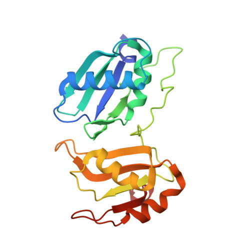
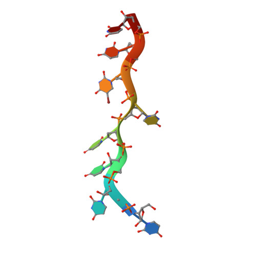
An extended U2AF(65)-RNA-binding domain recognizes the 3' splice site signal.

Agrawal, A.A.Salsi, E.Chatrikhi, R.Henderson, S.Jenkins, J.L.Green, M.R.Ermolenko, D.N.Kielkopf, C.L.

(2016) Nat Commun 7: 10950-10950

  • DOI: https://doi.org/10.1038/ncomms10950
  • Primary Citation of Related Structures:  
    5EV1, 5EV2, 5EV3, 5EV4

  • PubMed Abstract: 

    How the essential pre-mRNA splicing factor U2AF(65) recognizes the polypyrimidine (Py) signals of the major class of 3' splice sites in human gene transcripts remains incompletely understood. We determined four structures of an extended U2AF(65)-RNA-binding domain bound to Py-tract oligonucleotides at resolutions between 2.0 and 1.5 Å. These structures together with RNA binding and splicing assays reveal unforeseen roles for U2AF(65) inter-domain residues in recognizing a contiguous, nine-nucleotide Py tract. The U2AF(65) linker residues between the dual RNA recognition motifs (RRMs) recognize the central nucleotide, whereas the N- and C-terminal RRM extensions recognize the 3' terminus and third nucleotide. Single-molecule FRET experiments suggest that conformational selection and induced fit of the U2AF(65) RRMs are complementary mechanisms for Py-tract association. Altogether, these results advance the mechanistic understanding of molecular recognition for a major class of splice site signals.


  • Organizational Affiliation

    Center for RNA Biology and Department of Biochemistry and Biophysics, University of Rochester School of Medicine and Dentistry, Rochester, New York 14642, USA.


Macromolecules

Find similar proteins by:  (by identity cutoff)  |  3D Structure
Entity ID: 1
MoleculeChains Sequence LengthOrganismDetailsImage
Splicing factor U2AF 65 kDa subunit201Homo sapiensMutation(s): 0 
Gene Names: U2AF2U2AF65
UniProt & NIH Common Fund Data Resources
Find proteins for P26368 (Homo sapiens)
Explore P26368 
Go to UniProtKB:  P26368
PHAROS:  P26368
GTEx:  ENSG00000063244 
Entity Groups  
Sequence Clusters30% Identity50% Identity70% Identity90% Identity95% Identity100% Identity
UniProt GroupP26368
Sequence Annotations
Expand
  • Reference Sequence

Find similar nucleic acids by:  Sequence   |   3D Structure  

Entity ID: 2
MoleculeChains LengthOrganismImage
DNA/RNA (5'-R(*UP*UP*U)-D(P*UP*UP*(BRU)P*U)-R(P*UP*U)-3')9synthetic construct
Sequence Annotations
Expand
  • Reference Sequence
Experimental Data & Validation

Experimental Data

  • Method: X-RAY DIFFRACTION
  • Resolution: 2.04 Å
  • R-Value Free: 0.228 
  • R-Value Work: 0.173 
  • R-Value Observed: 0.178 
  • Space Group: C 2 2 21
Unit Cell:
Length ( Å )Angle ( ˚ )
a = 62.052α = 90
b = 114.241β = 90
c = 59.418γ = 90
Software Package:
Software NamePurpose
PHENIXrefinement
SCALAdata scaling
PDB_EXTRACTdata extraction
Blu-Icedata collection
XDSdata reduction

Structure Validation

View Full Validation Report


Entry History & Funding Information

Deposition Data


Funding OrganizationLocationGrant Number
National Institutes of Health/National Institute of General Medical Sciences (NIH/NIGMS)United StatesGM070503

Revision History  (Full details and data files)

  • Version 1.0: 2016-02-24
    Type: Initial release
  • Version 1.1: 2016-03-23
    Changes: Database references
  • Version 1.2: 2017-09-27
    Changes: Author supporting evidence, Derived calculations
  • Version 1.3: 2019-12-25
    Changes: Author supporting evidence
  • Version 1.4: 2023-09-27
    Changes: Data collection, Database references, Derived calculations, Refinement description