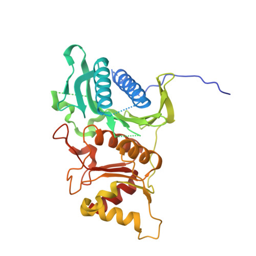
T-to-R switch of muscle fructose-1,6-bisphosphatase involves fundamental changes of secondary and quaternary structure.
Barciszewski, J., Wisniewski, J., Kolodziejczyk, R., Jaskolski, M., Rakus, D., Dzugaj, A.(2016) Acta Crystallogr D Struct Biol 72: 536-550
- PubMed: 27050133
- DOI: https://doi.org/10.1107/S2059798316001765
- Primary Citation of Related Structures:
5ET5, 5ET6, 5ET7 - PubMed Abstract:
Fructose-1,6-bisphosphatase (FBPase) catalyzes the hydrolysis of fructose 1,6-bisphosphate to fructose 6-phosphate and is a key enzyme of gluconeogenesis and glyconeogenesis and, more generally, of the control of energy metabolism and glucose homeostasis. Vertebrates, and notably Homo sapiens, express two FBPase isoforms. The liver isozyme is expressed mainly in gluconeogenic organs, where it functions as a regulator of glucose synthesis. The muscle isoform is expressed in all cells, and recent studies have demonstrated that its role goes far beyond the enzymatic function, as it can interact with various nuclear and mitochondrial proteins. Even in its enzymatic function, the muscle enzyme is different from the liver isoform, as it is 100-fold more susceptible to allosteric inhibition by AMP and this effect can be abrogated by complex formation with aldolase. All FBPases are homotetramers composed of two intimate dimers: the upper dimer and the lower dimer. They oscillate between two conformational states: the inactive T form when in complex with AMP, and the active R form. Parenthetically, it is noted that bacterial FBPases behave somewhat differently, and in the absence of allosteric activators exist in a tetramer-dimer equilibrium even at relatively high concentrations. [Hines et al. (2007), J. Biol. Chem. 282, 11696-11704]. The T-to-R transition is correlated with the conformation of the key loop L2, which in the T form becomes `disengaged' and unable to participate in the catalytic mechanism. The T states of both isoforms are very similar, with a small twist of the upper dimer relative to the lower dimer. It is shown that at variance with the well studied R form of the liver enzyme, which is flat, the R form of the muscle enzyme is diametrically different, with a perpendicular orientation of the upper and lower dimers. The crystal structure of the muscle-isozyme R form shows that in this arrangement of the tetramer completely new protein surfaces are exposed that are most likely targets for the interactions with various cellular and enzymatic partners. The cruciform R structure is stabilized by a novel `leucine lock', which prevents the key residue, Asp187, from locking loop L2 in the disengaged conformation. In addition, the crystal structures of muscle FBPase in the T conformation with and without AMP strongly suggest that the T-to-R transition is a discrete jump rather than a shift of an equilibrium smooth transition through multiple intermediate states. Finally, using snapshots from three crystal structures of human muscle FBPase, it is conclusively demonstrated that the AMP-binding event is correlated with a β→α transition at the N-terminus of the protein and with the formation of a new helical structure.
Organizational Affiliation:
Center for Biocrystallographic Research, Institute of Bioorganic Chemistry, Polish Academy of Sciences, Poznan, Poland.