5ELR

Structure of the KH-QUA2 domain of T-STAR in complex with AAUAAU RNA


Experimental Data Snapshot

  • Method: X-RAY DIFFRACTION
  • Resolution: 2.30 Å
  • R-Value Free: 0.269 
  • R-Value Work: 0.227 
  • R-Value Observed: 0.229 

wwPDB Validation   3D Report Full Report


This is version 1.2 of the entry. See complete history


Literature

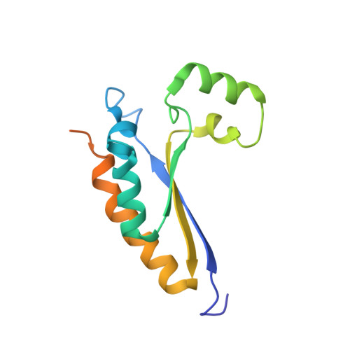
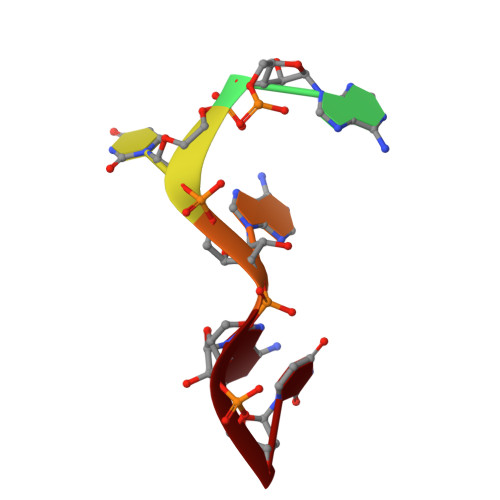
Structural basis of RNA recognition and dimerization by the STAR proteins T-STAR and Sam68.

Feracci, M.Foot, J.N.Grellscheid, S.N.Danilenko, M.Stehle, R.Gonchar, O.Kang, H.S.Dalgliesh, C.Meyer, N.H.Liu, Y.Lahat, A.Sattler, M.Eperon, I.C.Elliott, D.J.Dominguez, C.

(2016) Nat Commun 7: 10355-10355

  • DOI: https://doi.org/10.1038/ncomms10355
  • Primary Citation of Related Structures:  
    5EL3, 5ELR, 5ELS, 5ELT, 5EMO

  • PubMed Abstract: 

    Sam68 and T-STAR are members of the STAR family of proteins that directly link signal transduction with post-transcriptional gene regulation. Sam68 controls the alternative splicing of many oncogenic proteins. T-STAR is a tissue-specific paralogue that regulates the alternative splicing of neuronal pre-mRNAs. STAR proteins differ from most splicing factors, in that they contain a single RNA-binding domain. Their specificity of RNA recognition is thought to arise from their property to homodimerize, but how dimerization influences their function remains unknown. Here, we establish at atomic resolution how T-STAR and Sam68 bind to RNA, revealing an unexpected mode of dimerization different from other members of the STAR family. We further demonstrate that this unique dimerization interface is crucial for their biological activity in splicing regulation, and suggest that the increased RNA affinity through dimer formation is a crucial parameter enabling these proteins to select their functional targets within the transcriptome.


  • Organizational Affiliation

    Department of Molecular and Cell Biology, University of Leicester, Henry Wellcome Building, Lancaster Road, Leicester LE1 9HN, UK.


Macromolecules

Find similar proteins by:  (by identity cutoff)  |  3D Structure
Entity ID: 2
MoleculeChains Sequence LengthOrganismDetailsImage
KH domain-containing, RNA-binding, signal transduction-associated protein 3B [auth C],
C [auth D]
136Homo sapiensMutation(s): 0 
Gene Names: KHDRBS3SALPSLM2
UniProt & NIH Common Fund Data Resources
Find proteins for O75525 (Homo sapiens)
Explore O75525 
Go to UniProtKB:  O75525
PHAROS:  O75525
GTEx:  ENSG00000131773 
Entity Groups  
Sequence Clusters30% Identity50% Identity70% Identity90% Identity95% Identity100% Identity
UniProt GroupO75525
Sequence Annotations
Expand
  • Reference Sequence

Find similar nucleic acids by:  Sequence   |   3D Structure  

Entity ID: 1
MoleculeChains LengthOrganismImage
RNA (5'-R(*AP*AP*UP*AP*AP*U)-3')A [auth B]6synthetic construct
Sequence Annotations
Expand
  • Reference Sequence
Experimental Data & Validation

Experimental Data

  • Method: X-RAY DIFFRACTION
  • Resolution: 2.30 Å
  • R-Value Free: 0.269 
  • R-Value Work: 0.227 
  • R-Value Observed: 0.229 
  • Space Group: P 21 21 21
Unit Cell:
Length ( Å )Angle ( ˚ )
a = 42.38α = 90
b = 45.56β = 90
c = 151.98γ = 90
Software Package:
Software NamePurpose
Aimlessdata scaling
REFMACrefinement
PDB_EXTRACTdata extraction
PHASERphasing
xia2data reduction

Structure Validation

View Full Validation Report


Entry History & Funding Information

Deposition Data


Funding OrganizationLocationGrant Number
Medical Research Council (United Kingdom)United KingdomG1000526

Revision History  (Full details and data files)

  • Version 1.0: 2016-01-13
    Type: Initial release
  • Version 1.1: 2016-01-27
    Changes: Database references
  • Version 1.2: 2017-09-13
    Changes: Author supporting evidence, Data collection