Binding and structural studies of the complexes of type 1 ribosome inactivating protein fromMomordica balsaminawith cytosine, cytidine, and cytidine diphosphate.
Yamini, S., Pandey, S.N., Kaur, P., Sharma, S., Singh, T.P.(2015) Biochem Biophys Rep 4: 134-140
- PubMed: 29124196
- DOI: https://doi.org/10.1016/j.bbrep.2015.09.006
- Primary Citation of Related Structures:
4ZT8, 4ZU0, 4ZZ6, 5CSO, 5CST - PubMed Abstract:
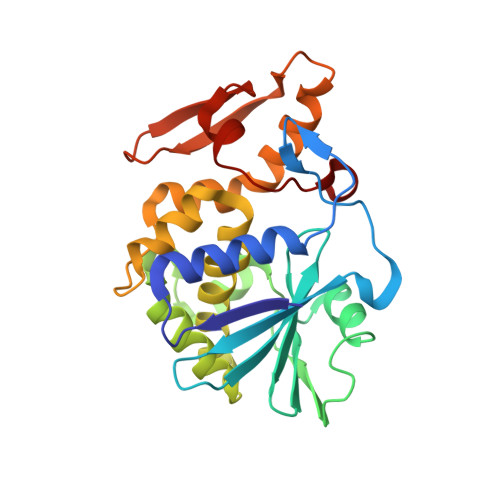
The type 1 ribosome inactivating protein from Momordica balsamina ( Mb RIP1) has been shown to interact with purine bases, adenine and guanine of RNA/DNA. We report here the binding and structural studies of Mb RIP1 with a pyrimidine base, cytosine; cytosine containing nucleoside, cytidine; and cytosine containing nucleotide, cytidine diphosphate. All three compounds bound to Mb RIP1 at the active site with dissociation constants of 10 -4 M-10 -7 M. As reported earlier, in the structure of native Mb RIP1, there are 10 water molecules in the substrate binding site. Upon binding of cytosine to Mb RIP1, four water molecules were dislodged from the substrate binding site while five water molecules were dislodged when cytidine bound to Mb RIP1. Seven water molecules were dislocated when cytidine diphosphate bound to Mb RIP1. This showed that cytidine diphosphate occupied a larger space in the substrate binding site enhancing the buried surface area thus making it a relatively better inhibitor of Mb RIP1 as compared to cytosine and cytidine. The key residues involved in the recognition of cytosine, cytidine and cytidine diphosphate were Ile71, Glu85, Tyr111 and Arg163. The orientation of cytosine in the cleft is different from that of adenine or guanine indicating a notable difference in the modes of binding of purine and pyrimidine bases. Since adenine containing nucleosides/nucleotides are suitable substrates, the cytosine containing nucleosides/nucleotides may act as inhibitors.
Organizational Affiliation:
Department of Biophysics, All India institute of Medical Sciences, New Delhi, India.