5CPH

Crystal structure of the ATP binding domain of S. aureus GyrB complexed with a fragment


Experimental Data Snapshot

  • Method: X-RAY DIFFRACTION
  • Resolution: 1.20 Å
  • R-Value Free: 0.194 
  • R-Value Work: 0.172 
  • R-Value Observed: 0.173 

wwPDB Validation   3D Report Full Report


Ligand Structure Quality Assessment 


This is version 1.2 of the entry. See complete history


Literature

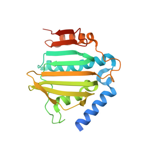
Fragment-based discovery of DNA gyrase inhibitors targeting the ATPase subunit of GyrB.

Mesleh, M.F.Cross, J.B.Zhang, J.Kahmann, J.Andersen, O.A.Barker, J.Cheng, R.K.Felicetti, B.Wood, M.Hadfield, A.T.Scheich, C.Moy, T.I.Yang, Q.Shotwell, J.Nguyen, K.Lippa, B.Dolle, R.Ryan, M.D.

(2016) Bioorg Med Chem Lett 26: 1314-1318

  • DOI: https://doi.org/10.1016/j.bmcl.2016.01.009
  • Primary Citation of Related Structures:  
    5CPH, 5CTU, 5CTW, 5CTX, 5CTY

  • PubMed Abstract: 

    Inhibitors of the ATPase function of bacterial DNA gyrase, located in the GyrB subunit and its related ParE subunit in topoisomerase IV, have demonstrated antibacterial activity. In this study we describe an NMR fragment-based screening effort targeting Staphylococcus aureus GyrB that identified several attractive and novel starting points with good ligand efficiency. Fragment hits were further characterized using NMR binding studies against full-length S. aureus GyrB and Escherichia coli ParE. X-ray co-crystal structures of select fragment hits confirmed binding and suggested a path for medicinal chemistry optimization. The identification, characterization, and elaboration of one of these fragment series to a 0.265 μM inhibitor is described herein.


  • Organizational Affiliation

    Cubist Pharmaceuticals, 65 Hayden Ave., Lexington, MA 02421, United States.


Macromolecules
Find similar proteins by:  (by identity cutoff)  |  3D Structure
Entity ID: 1
MoleculeChains Sequence LengthOrganismDetailsImage
DNA gyrase subunit B
A, B
212Staphylococcus aureusMutation(s): 0 
Gene Names: gyrB
EC: 5.99.1.3
UniProt
Find proteins for P0A0K8 (Staphylococcus aureus)
Explore P0A0K8 
Go to UniProtKB:  P0A0K8
Entity Groups  
Sequence Clusters30% Identity50% Identity70% Identity90% Identity95% Identity100% Identity
UniProt GroupP0A0K8
Sequence Annotations
Expand
  • Reference Sequence
Small Molecules
Binding Affinity Annotations 
IDSourceBinding Affinity
EVO BindingDB:  5CPH IC50: 8.40e+5 (nM) from 1 assay(s)
Experimental Data & Validation

Experimental Data

  • Method: X-RAY DIFFRACTION
  • Resolution: 1.20 Å
  • R-Value Free: 0.194 
  • R-Value Work: 0.172 
  • R-Value Observed: 0.173 
  • Space Group: C 1 2 1
Unit Cell:
Length ( Å )Angle ( ˚ )
a = 142.852α = 90
b = 55.681β = 100.69
c = 51.569γ = 90
Software Package:
Software NamePurpose
REFMACrefinement
d*TREKdata reduction
d*TREKdata scaling
MOLREPphasing
PDB_EXTRACTdata extraction

Structure Validation

View Full Validation Report


Ligand Structure Quality Assessment 


Entry History 

Deposition Data

Revision History  (Full details and data files)

  • Version 1.0: 2016-02-03
    Type: Initial release
  • Version 1.1: 2016-02-17
    Changes: Structure summary
  • Version 1.2: 2023-09-27
    Changes: Data collection, Database references, Derived calculations, Refinement description