


Crystal structure of a novel disulfide oxidoreductase from Deinococcus radiodurans
Bihani, S.C., Panicker, L., Kumar, V., Misra, H.S., Rajpurohit, Y.S.To be published.
Experimental Data Snapshot
wwPDB Validation 3D Report Full Report

Entity ID: 1 | |||||
|---|---|---|---|---|---|
| Molecule | Chains | Sequence Length | Organism | Details | Image |
| FrnE protein | 260 | Deinococcus radiodurans R1 = ATCC 13939 = DSM 20539 | Mutation(s): 1 Gene Names: DR_0659 | 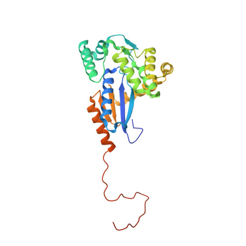 | |
UniProt | |||||
Find proteins for Q9RWK7 (Deinococcus radiodurans (strain ATCC 13939 / DSM 20539 / JCM 16871 / CCUG 27074 / LMG 4051 / NBRC 15346 / NCIMB 9279 / VKM B-1422 / R1)) Explore Q9RWK7 Go to UniProtKB: Q9RWK7 | |||||
Entity Groups | |||||
| Sequence Clusters | 30% Identity50% Identity70% Identity90% Identity95% Identity100% Identity | ||||
| UniProt Group | Q9RWK7 | ||||
Sequence AnnotationsExpand | |||||
| |||||
| Ligands 3 Unique | |||||
|---|---|---|---|---|---|
| ID | Chains | Name / Formula / InChI Key | 2D Diagram | 3D Interactions | |
| GOL Query on GOL | D [auth A], E [auth A], F [auth A], G [auth A] | GLYCEROL C3 H8 O3 PEDCQBHIVMGVHV-UHFFFAOYSA-N |  | ||
| EDO Query on EDO | H [auth A] | 1,2-ETHANEDIOL C2 H6 O2 LYCAIKOWRPUZTN-UHFFFAOYSA-N |  | ||
| ACT Query on ACT | B [auth A], C [auth A] | ACETATE ION C2 H3 O2 QTBSBXVTEAMEQO-UHFFFAOYSA-M |  | ||
| Length ( Å ) | Angle ( ˚ ) |
|---|---|
| a = 49.091 | α = 90 |
| b = 66.158 | β = 90 |
| c = 84.095 | γ = 90 |
| Software Name | Purpose |
|---|---|
| PHENIX | refinement |
| PDB_EXTRACT | data extraction |
| XDS | data processing |
| Aimless | data scaling |
| PHASER | phasing |
| XDS | data reduction |
RCSB PDB (citation) is hosted by
RCSB PDB is a member of the

