


Crystal Structure of the I80Y/L114V/I116V mutant of LEH
Wu, L., Sun, Z.T., Reetz, M.T., Zhou, J.H.To be published.
Experimental Data Snapshot
wwPDB Validation 3D Report Full Report

Entity ID: 1 | |||||
|---|---|---|---|---|---|
| Molecule | Chains | Sequence Length | Organism | Details | Image |
| Limonene-1,2-epoxide hydrolase | 155 | Rhodococcus erythropolis | Mutation(s): 3 Gene Names: limA EC: 3.3.2.8 | 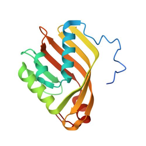 | |
UniProt | |||||
Find proteins for Q9ZAG3 (Rhodococcus erythropolis) Explore Q9ZAG3 Go to UniProtKB: Q9ZAG3 | |||||
Entity Groups | |||||
| Sequence Clusters | 30% Identity50% Identity70% Identity90% Identity95% Identity100% Identity | ||||
| UniProt Group | Q9ZAG3 | ||||
Sequence AnnotationsExpand | |||||
| |||||
| Length ( Å ) | Angle ( ˚ ) |
|---|---|
| a = 103.651 | α = 90 |
| b = 103.651 | β = 90 |
| c = 73.263 | γ = 120 |
| Software Name | Purpose |
|---|---|
| PHENIX | refinement |
| HKL-2000 | data reduction |
| SCALEPACK | data scaling |
| PHASER | phasing |
RCSB PDB (citation) is hosted by
RCSB PDB is a member of the

