5CA8

Structures of the yeast dynamin-like GTPase Sey1p in complex with GDP


Experimental Data Snapshot

  • Method: X-RAY DIFFRACTION
  • Resolution: 2.30 Å
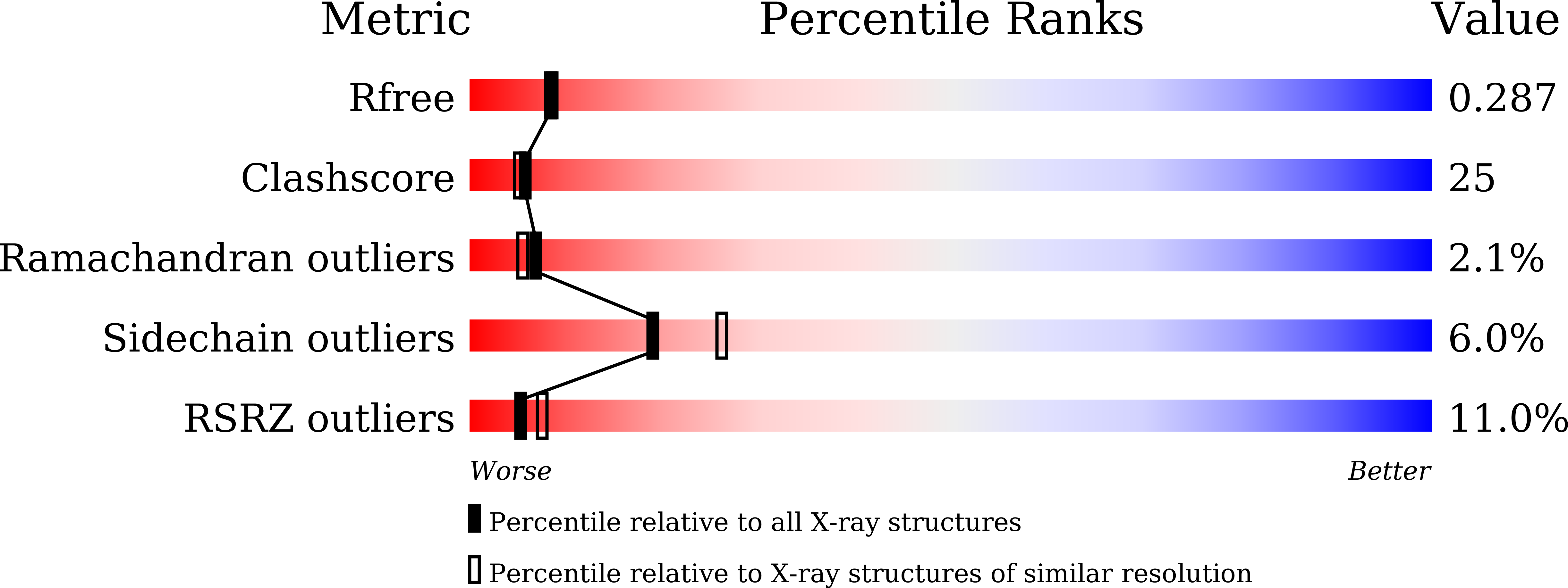
  • R-Value Free: 0.285 
  • R-Value Work: 0.230 
  • R-Value Observed: 0.233 

wwPDB Validation   3D Report Full Report


Ligand Structure Quality Assessment 


This is version 1.3 of the entry. See complete history


Literature

Structures of the yeast dynamin-like GTPase Sey1p provide insight into homotypic ER fusion

Yan, L.Sun, S.Wang, W.Shi, J.Hu, X.Wang, S.Su, D.Rao, Z.Hu, J.Lou, Z.

(2015) J Cell Biol 210: 961-972

  • DOI: https://doi.org/10.1083/jcb.201502078
  • Primary Citation of Related Structures:  
    5CA8, 5CA9, 5CB2

  • PubMed Abstract: 

    Homotypic membrane fusion of the endoplasmic reticulum is mediated by dynamin-like guanosine triphosphatases (GTPases), which include atlastin (ATL) in metazoans and Sey1p in yeast. In this paper, we determined the crystal structures of the cytosolic domain of Sey1p derived from Candida albicans. The structures reveal a stalk-like, helical bundle domain following the GTPase, which represents a previously unidentified configuration of the dynamin superfamily. This domain is significantly longer than that of ATL and critical for fusion. Sey1p forms a side-by-side dimer in complex with GMP-PNP or GDP/AlF4(-) but is monomeric with GDP. Surprisingly, Sey1p could mediate fusion without GTP hydrolysis, even though fusion was much more efficient with GTP. Sey1p was able to replace ATL in mammalian cells, and the punctate localization of Sey1p was dependent on its GTPase activity. Despite the common function of fusogenic GTPases, our results reveal unique features of Sey1p.


  • Organizational Affiliation

    School of Medicine, Tsinghua University, Beijing, China 100084.


Macromolecules
Find similar proteins by:  (by identity cutoff)  |  3D Structure
Entity ID: 1
MoleculeChains Sequence LengthOrganismDetailsImage
Protein SEY1692Candida albicans SC5314Mutation(s): 0 
Gene Names: SEY1NAG6CaO19.2151CaO19.9698
EC: 3.6.5.5
UniProt
Find proteins for Q9C0L9 (Candida albicans (strain SC5314 / ATCC MYA-2876))
Explore Q9C0L9 
Go to UniProtKB:  Q9C0L9
Entity Groups  
Sequence Clusters30% Identity50% Identity70% Identity90% Identity95% Identity100% Identity
UniProt GroupQ9C0L9
Sequence Annotations
Expand
  • Reference Sequence
Small Molecules
Ligands 2 Unique
IDChains Name / Formula / InChI Key2D Diagram3D Interactions
GDP
Query on GDP

Download Ideal Coordinates CCD File 
C [auth A]GUANOSINE-5'-DIPHOSPHATE
C10 H15 N5 O11 P2
QGWNDRXFNXRZMB-UUOKFMHZSA-N
MG
Query on MG

Download Ideal Coordinates CCD File 
B [auth A]MAGNESIUM ION
Mg
JLVVSXFLKOJNIY-UHFFFAOYSA-N
Experimental Data & Validation

Experimental Data

  • Method: X-RAY DIFFRACTION
  • Resolution: 2.30 Å
  • R-Value Free: 0.285 
  • R-Value Work: 0.230 
  • R-Value Observed: 0.233 
  • Space Group: C 1 2 1
Unit Cell:
Length ( Å )Angle ( ˚ )
a = 228.12α = 90
b = 43.62β = 108.12
c = 97.56γ = 90
Software Package:
Software NamePurpose
PHENIXrefinement
HKL-2000data processing
PHENIXmodel building
PHASERphasing

Structure Validation

View Full Validation Report


Ligand Structure Quality Assessment 


Entry History & Funding Information

Deposition Data

  • Released Date: 2015-09-16 
  • Deposition Author(s): Yan, L.

Funding OrganizationLocationGrant Number
National Natural Science FoundationChina81322023
973 ProgramChina2013CB911103

Revision History  (Full details and data files)

  • Version 1.0: 2015-09-16
    Type: Initial release
  • Version 1.1: 2015-10-07
    Changes: Database references
  • Version 1.2: 2020-02-19
    Changes: Data collection, Database references, Derived calculations
  • Version 1.3: 2023-11-08
    Changes: Data collection, Database references, Derived calculations, Refinement description