5BUA

Lysine 120-acetylated P53 DNA binding domain in a complex with DNA.


Experimental Data Snapshot

  • Method: X-RAY DIFFRACTION
  • Resolution: 1.81 Å
  • R-Value Free: 0.213 
  • R-Value Work: 0.162 
  • R-Value Observed: 0.165 

wwPDB Validation   3D Report Full Report


This is version 1.2 of the entry. See complete history


Literature

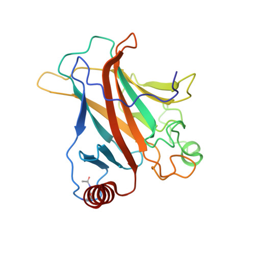
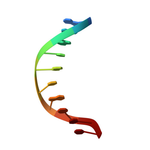
Structural Basis for p53 Lys120-Acetylation-Dependent DNA-Binding Mode.

Vainer, R.Cohen, S.Shahar, A.Zarivach, R.Arbely, E.

(2016) J Mol Biol 428: 3013-3025

  • DOI: https://doi.org/10.1016/j.jmb.2016.06.009
  • Primary Citation of Related Structures:  
    5BUA, 5LGY

  • PubMed Abstract: 

    Normal cellular homeostasis depends on tight regulation of gene expression, which requires the modulation of transcription factors' DNA-binding specificity. That said, the mechanisms that allow transcription factors to distinguish between closely related response elements following different cellular signals are not fully understood. In the tumor suppressor protein p53, acetylation of loop L1 residue Lys120 within the DNA-binding domain has been shown to promote the transcription of proapoptotic genes such as bax. Here, we report the crystal structures of Lys120-acetylated p53 DNA-binding domain in complex with a consensus response element and with the natural BAX response element. Our structural analyses reveal that Lys120 acetylation expands the conformational space of loop L1 in the DNA-bound state. Loop L1 flexibility is known to increase p53's DNA-binding specificity, and Lys120-acetylation-dependent conformational changes in loop L1 enable the formation of sequence-dependent DNA-binding modes for p53. Furthermore, binding to the natural BAX response element is accompanied by global conformational changes, deformation of the DNA helical structure, and formation of an asymmetric tetrameric complex. Based on these findings, we suggest a model for p53's Lys120 acetylation-dependent DNA-binding mode.


  • Organizational Affiliation

    Department of Chemistry, Ben-Gurion University of the Negev, Beer-Sheva, 84105, Israel.


Macromolecules

Find similar proteins by:  (by identity cutoff)  |  3D Structure
Entity ID: 1
MoleculeChains Sequence LengthOrganismDetailsImage
Cellular tumor antigen p53200Homo sapiensMutation(s): 0 
Gene Names: TP53P53
UniProt & NIH Common Fund Data Resources
Find proteins for P04637 (Homo sapiens)
Explore P04637 
Go to UniProtKB:  P04637
PHAROS:  P04637
GTEx:  ENSG00000141510 
Entity Groups  
Sequence Clusters30% Identity50% Identity70% Identity90% Identity95% Identity100% Identity
UniProt GroupP04637
Sequence Annotations
Expand
  • Reference Sequence

Find similar nucleic acids by:  Sequence   |   3D Structure  

Entity ID: 2
MoleculeChains LengthOrganismImage
DNA (5'-D(P*GP*GP*AP*CP*AP*TP*GP*TP*CP*C)-3')10synthetic construct
Sequence Annotations
Expand
  • Reference Sequence
Small Molecules
Ligands 1 Unique
IDChains Name / Formula / InChI Key2D Diagram3D Interactions
ZN
Query on ZN

Download Ideal Coordinates CCD File 
C [auth A]ZINC ION
Zn
PTFCDOFLOPIGGS-UHFFFAOYSA-N
Modified Residues  1 Unique
IDChains TypeFormula2D DiagramParent
ALY
Query on ALY
A
L-PEPTIDE LINKINGC8 H16 N2 O3LYS
Experimental Data & Validation

Experimental Data

  • Method: X-RAY DIFFRACTION
  • Resolution: 1.81 Å
  • R-Value Free: 0.213 
  • R-Value Work: 0.162 
  • R-Value Observed: 0.165 
  • Space Group: C 1 2 1
Unit Cell:
Length ( Å )Angle ( ˚ )
a = 137.252α = 90
b = 48.892β = 92.6
c = 34.014γ = 90
Software Package:
Software NamePurpose
PHENIXrefinement
DENZOdata reduction
SCALEPACKdata scaling
PHASERphasing
PDB_EXTRACTdata extraction

Structure Validation

View Full Validation Report


Entry History & Funding Information

Deposition Data


Funding OrganizationLocationGrant Number
Concern FoundationUnited States--

Revision History  (Full details and data files)

  • Version 1.0: 2016-07-06
    Type: Initial release
  • Version 1.1: 2016-08-17
    Changes: Database references
  • Version 1.2: 2024-01-10
    Changes: Data collection, Database references, Refinement description