5B3I

Homo-dimeric structure of cytochrome c' from Thermophilic Hydrogenophilus thermoluteolus


Experimental Data Snapshot

  • Method: X-RAY DIFFRACTION
  • Resolution: 1.89 Å
  • R-Value Free: 0.243 
  • R-Value Work: 0.196 
  • R-Value Observed: 0.198 

wwPDB Validation   3D Report Full Report


Ligand Structure Quality Assessment 


This is version 2.1 of the entry. See complete history


Literature

Structural and functional insights into thermally stable cytochrome c' from a thermophile

Fujii, S.Oki, H.Kawahara, K.Yamane, D.Yamanaka, M.Maruno, T.Kobayashi, Y.Masanari, M.Wakai, S.Nishihara, H.Ohkubo, T.Sambongi, Y.

(2017) Protein Sci 26: 737-748

  • DOI: https://doi.org/10.1002/pro.3120
  • Primary Citation of Related Structures:  
    5B3I

  • PubMed Abstract: 

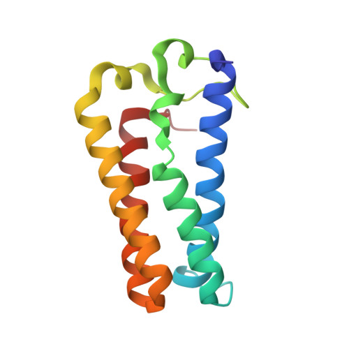
    Thermophilic Hydrogenophilus thermoluteolus cytochrome c' (PHCP) exhibits higher thermal stability than a mesophilic counterpart, Allochromatium vinosum cytochrome c' (AVCP), which has a homo-dimeric structure and ligand-binding ability. To understand the thermal stability mechanism and ligand-binding ability of the thermally stable PHCP protein, the crystal structure of PHCP was first determined. It formed a homo-dimeric structure, the main chain root mean square deviation (rmsd) value between PHCP and AVCP being 0.65 Å. In the PHCP structure, six specific residues appeared to strengthen the heme-related and subunit-subunit interactions, which were not conserved in the AVCP structure. PHCP variants having altered subunit-subunit interactions were more severely destabilized than ones having altered heme-related interactions. The PHCP structure further revealed a ligand-binding channel and a penta-coordinated heme, as observed in the AVCP protein. A spectroscopic study clearly showed that some ligands were bound to the PHCP protein. It is concluded that the dimeric PHCP from the thermophile is effectively stabilized through heme-related and subunit-subunit interactions with conservation of the ligand-binding ability.


  • Organizational Affiliation

    Graduate School of Biosphere Science, Hiroshima University, Higashi-Hiroshima, Hiroshima, Japan.


Macromolecules
Find similar proteins by:  (by identity cutoff)  |  3D Structure
Entity ID: 1
MoleculeChains Sequence LengthOrganismDetailsImage
Cytochrome c prime
A, B, C, D
135Hydrogenophilus thermoluteolusMutation(s): 0 
UniProt
Find proteins for F7J213 (Hydrogenophilus thermoluteolus)
Explore F7J213 
Go to UniProtKB:  F7J213
Entity Groups  
Sequence Clusters30% Identity50% Identity70% Identity90% Identity95% Identity100% Identity
UniProt GroupF7J213
Sequence Annotations
Expand
  • Reference Sequence
Experimental Data & Validation

Experimental Data

  • Method: X-RAY DIFFRACTION
  • Resolution: 1.89 Å
  • R-Value Free: 0.243 
  • R-Value Work: 0.196 
  • R-Value Observed: 0.198 
  • Space Group: P 21 2 21
Unit Cell:
Length ( Å )Angle ( ˚ )
a = 54.243α = 90
b = 72.923β = 90
c = 132.365γ = 90
Software Package:
Software NamePurpose
PHENIXrefinement
CrystalCleardata collection
d*TREKdata reduction
d*TREKdata scaling
PHASERphasing
PDB_EXTRACTdata extraction

Structure Validation

View Full Validation Report


Ligand Structure Quality Assessment 


Entry History 

Deposition Data

Revision History  (Full details and data files)

  • Version 1.0: 2017-03-01
    Type: Initial release
  • Version 1.1: 2017-04-05
    Changes: Database references
  • Version 2.0: 2019-10-02
    Changes: Advisory, Atomic model, Data collection, Derived calculations, Non-polymer description, Structure summary
  • Version 2.1: 2023-11-08
    Changes: Data collection, Database references, Derived calculations, Refinement description