5B0T

Structure of Shigella effector LRR domain


Experimental Data Snapshot

  • Method: X-RAY DIFFRACTION
  • Resolution: 2.00 Å
  • R-Value Free: 0.265 
  • R-Value Work: 0.211 
  • R-Value Observed: 0.214 

wwPDB Validation   3D Report Full Report


This is version 1.3 of the entry. See complete history


Literature

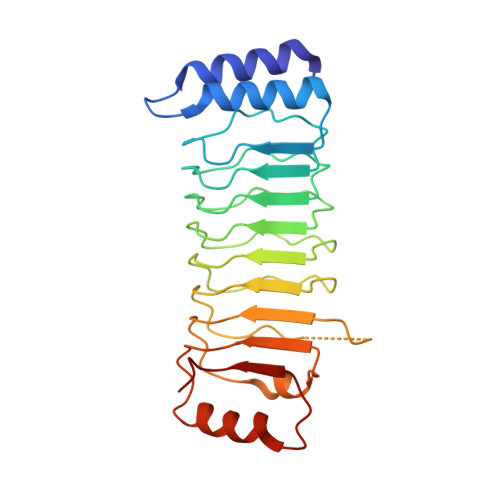
Crystal structure of the substrate-recognition domain of the Shigella E3 ligase IpaH9.8

Takagi, K.Kim, M.Sasakawa, C.Mizushima, T.

(2016) Acta Crystallogr F Struct Biol Commun 72: 269-275

  • DOI: https://doi.org/10.1107/S2053230X16002715
  • Primary Citation of Related Structures:  
    5B0N, 5B0T

  • PubMed Abstract: 

    Infectious diseases caused by bacteria have significant impacts on global public health. During infection, pathogenic bacteria deliver a variety of virulence factors, called effectors, into host cells. The Shigella effector IpaH9.8 functions as an ubiquitin ligase, ubiquitinating the NF-κB essential modulator (NEMO)/IKK-γ to inhibit host inflammatory responses. IpaH9.8 contains leucine-rich repeats (LRRs) involved in substrate recognition and an E3 ligase domain. To elucidate the structural basis of the function of IpaH9.8, the crystal structure of the LRR domain of Shigella IpaH9.8 was determined and this structure was compared with the known structures of other IpaH family members. This model provides insights into the structural features involved in substrate specificity.


  • Organizational Affiliation

    Graduate School of Life Science, University of Hyogo, 3-2-1 Kouto, Kamigori-cho, Ako-gun, Hyogo 678-1297, Japan.


Macromolecules
Find similar proteins by:  (by identity cutoff)  |  3D Structure
Entity ID: 1
MoleculeChains Sequence LengthOrganismDetailsImage
E3 ubiquitin-protein ligase ipaH9.8224Shigella flexneriMutation(s): 0 
Gene Names: ipaH9.8CP0226pWR501_0234SFLP090
EC: 6.3.2
UniProt
Find proteins for Q8VSC3 (Shigella flexneri)
Explore Q8VSC3 
Go to UniProtKB:  Q8VSC3
Entity Groups  
Sequence Clusters30% Identity50% Identity70% Identity90% Identity95% Identity100% Identity
UniProt GroupQ8VSC3
Sequence Annotations
Expand
  • Reference Sequence
Experimental Data & Validation

Experimental Data

  • Method: X-RAY DIFFRACTION
  • Resolution: 2.00 Å
  • R-Value Free: 0.265 
  • R-Value Work: 0.211 
  • R-Value Observed: 0.214 
  • Space Group: C 2 2 21
Unit Cell:
Length ( Å )Angle ( ˚ )
a = 68.271α = 90
b = 105.011β = 90
c = 61.596γ = 90
Software Package:
Software NamePurpose
REFMACrefinement
HKL-2000data collection
HKL-2000data scaling
MOLREPphasing
PDB_EXTRACTdata extraction

Structure Validation

View Full Validation Report


Entry History 

Deposition Data

Revision History  (Full details and data files)

  • Version 1.0: 2016-04-06
    Type: Initial release
  • Version 1.1: 2016-04-20
    Changes: Database references
  • Version 1.2: 2020-02-26
    Changes: Data collection, Derived calculations
  • Version 1.3: 2024-03-20
    Changes: Data collection, Database references