5AJT

Crystal structure of ligand-free phosphoribohydrolase lonely guy from Claviceps purpurea


Experimental Data Snapshot

  • Method: X-RAY DIFFRACTION
  • Resolution: 2.43 Å
  • R-Value Free: 0.240 
  • R-Value Work: 0.215 
  • R-Value Observed: 0.216 

wwPDB Validation   3D Report Full Report


This is version 2.1 of the entry. See complete history


Literature

The three-dimensional structure of "Lonely Guy" from Claviceps purpurea provides insights into the phosphoribohydrolase function of Rossmann fold-containing lysine decarboxylase-like proteins.

Dzurova, L.Forneris, F.Savino, S.Galuszka, P.Vrabka, J.Frebort, I.

(2015) Proteins 83: 1539-1546

  • DOI: https://doi.org/10.1002/prot.24835
  • Primary Citation of Related Structures:  
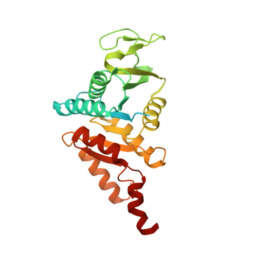
    5AJT

  • PubMed Abstract: 

    The recently discovered cytokinin (CK)-specific phosphoribohydrolase "Lonely Guy" (LOG) is a key enzyme of CK biosynthesis, converting inactive CK nucleotides into biologically active free bases. We have determined the crystal structures of LOG from Claviceps purpurea (cpLOG) and its complex with the enzymatic product phosphoribose. The structures reveal a dimeric arrangement of Rossmann folds, with the ligands bound to large pockets at the interface between cpLOG monomers. Structural comparisons highlight the homology of cpLOG to putative lysine decarboxylases. Extended sequence analysis enabled identification of a distinguishing LOG sequence signature. Taken together, our data suggest phosphoribohydrolase activity for several proteins of unknown function.


  • Organizational Affiliation

    Centre of the Region Haná for Biotechnological and Agricultural Research, Faculty of Science, Palacký University, Olomouc, 783 71, Czech Republic.


Macromolecules
Find similar proteins by:  (by identity cutoff)  |  3D Structure
Entity ID: 1
MoleculeChains Sequence LengthOrganismDetailsImage
PHOSPHORIBOHYDROLASE LONELY GUY
A, B, C, D
274Claviceps purpureaMutation(s): 0 
UniProt
Find proteins for M1VUY5 (Claviceps purpurea (strain 20.1))
Explore M1VUY5 
Go to UniProtKB:  M1VUY5
Entity Groups  
Sequence Clusters30% Identity50% Identity70% Identity90% Identity95% Identity100% Identity
UniProt GroupM1VUY5
Sequence Annotations
Expand
  • Reference Sequence
Small Molecules
Ligands 2 Unique
IDChains Name / Formula / InChI Key2D Diagram3D Interactions
TAR
Query on TAR

Download Ideal Coordinates CCD File 
G [auth A],
I [auth B],
K [auth C],
M [auth D],
N [auth D]
D(-)-TARTARIC ACID
C4 H6 O6
FEWJPZIEWOKRBE-LWMBPPNESA-N
EDO
Query on EDO

Download Ideal Coordinates CCD File 
E [auth A],
F [auth A],
H [auth B],
J [auth C],
L [auth D]
1,2-ETHANEDIOL
C2 H6 O2
LYCAIKOWRPUZTN-UHFFFAOYSA-N
Experimental Data & Validation

Experimental Data

  • Method: X-RAY DIFFRACTION
  • Resolution: 2.43 Å
  • R-Value Free: 0.240 
  • R-Value Work: 0.215 
  • R-Value Observed: 0.216 
  • Space Group: I 1 2 1
Unit Cell:
Length ( Å )Angle ( ˚ )
a = 82.54α = 90
b = 83.17β = 94.03
c = 163.652γ = 90
Software Package:
Software NamePurpose
PHENIXrefinement
iMOSFLMdata reduction
Aimlessdata scaling
PHASERphasing

Structure Validation

View Full Validation Report


Entry History 

Deposition Data

Revision History  (Full details and data files)

  • Version 1.0: 2015-06-03
    Type: Initial release
  • Version 1.1: 2015-08-05
    Changes: Database references
  • Version 2.0: 2019-06-26
    Changes: Atomic model, Data collection, Database references, Derived calculations, Experimental preparation, Non-polymer description, Other, Structure summary
  • Version 2.1: 2024-01-10
    Changes: Data collection, Database references, Derived calculations, Other, Refinement description