5AG2

SOD-3 azide complex


Experimental Data Snapshot

  • Method: X-RAY DIFFRACTION
  • Resolution: 1.77 Å
  • R-Value Free: 0.216 
  • R-Value Work: 0.185 
  • R-Value Observed: 0.186 

wwPDB Validation   3D Report Full Report


This is version 1.1 of the entry. See complete history


Literature

The Structure of the Caenorhabditis Elegans Manganese Superoxide Dismutase Mnsod-3-Azide Complex.

Hunter, G.J.Trinh, C.H.Bonetta, R.Stewart, E.E.Cabelli, D.E.Hunter, T.

(2015) Protein Sci 24: 1777

  • DOI: https://doi.org/10.1002/pro.2768
  • Primary Citation of Related Structures:  
    4X9Q, 5AG2

  • PubMed Abstract: 

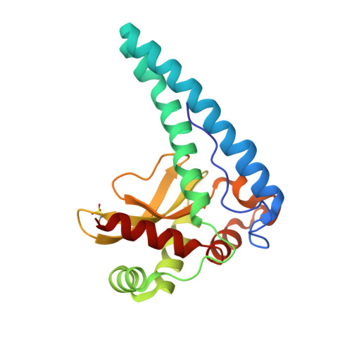
    C. elegans MnSOD-3 has been implicated in the longevity pathway and its mechanism of catalysis is relevant to the aging process and carcinogenesis. The structures of MnSOD-3 provide unique crystallographic evidence of a dynamic region of the tetrameric interface (residues 41-54). We have determined the structure of the MnSOD-3-azide complex to 1.77-Å resolution. Analysis of this complex shows that the substrate analog, azide, binds end-on to the manganese center as a sixth ligand and that it ligates directly to a third and new solvent molecule also positioned within interacting distance to the His30 and Tyr34 residues of the substrate access funnel. This is the first structure of a eukaryotic MnSOD-azide complex that demonstrates the extended, uninterrupted hydrogen-bonded network that forms a proton relay incorporating three outer sphere solvent molecules, the substrate analog, the gateway residues, Gln142, and the solvent ligand. This configuration supports the formation and release of the hydrogen peroxide product in agreement with the 5-6-5 catalytic mechanism for MnSOD. The high product dissociation constant k4 of MnSOD-3 reflects low product inhibition making this enzyme efficient even at high levels of superoxide.


  • Organizational Affiliation

    Department of Physiology and Biochemistry, Faculty of Medicine and Surgery, University of Malta, Msida, Malta.


Macromolecules
Find similar proteins by:  (by identity cutoff)  |  3D Structure
Entity ID: 1
MoleculeChains Sequence LengthOrganismDetailsImage
SUPEROXIDE DISMUTASE [MN] 2, MITOCHONDRIALA,
B [auth C]
195Caenorhabditis elegansMutation(s): 0 
EC: 1.15.1.1
UniProt
Find proteins for P41977 (Caenorhabditis elegans)
Explore P41977 
Go to UniProtKB:  P41977
Entity Groups  
Sequence Clusters30% Identity50% Identity70% Identity90% Identity95% Identity100% Identity
UniProt GroupP41977
Sequence Annotations
Expand
  • Reference Sequence
Small Molecules
Ligands 5 Unique
IDChains Name / Formula / InChI Key2D Diagram3D Interactions
MLI
Query on MLI

Download Ideal Coordinates CCD File 
F [auth A]MALONATE ION
C3 H2 O4
OFOBLEOULBTSOW-UHFFFAOYSA-L
SO4
Query on SO4

Download Ideal Coordinates CCD File 
I [auth C]SULFATE ION
O4 S
QAOWNCQODCNURD-UHFFFAOYSA-L
ACT
Query on ACT

Download Ideal Coordinates CCD File 
E [auth A]ACETATE ION
C2 H3 O2
QTBSBXVTEAMEQO-UHFFFAOYSA-M
MN
Query on MN

Download Ideal Coordinates CCD File 
C [auth A],
G [auth C]
MANGANESE (II) ION
Mn
WAEMQWOKJMHJLA-UHFFFAOYSA-N
AZI
Query on AZI

Download Ideal Coordinates CCD File 
D [auth A],
H [auth C]
AZIDE ION
N3
IVRMZWNICZWHMI-UHFFFAOYSA-N
Modified Residues  1 Unique
IDChains TypeFormula2D DiagramParent
OCS
Query on OCS
A,
B [auth C]
L-PEPTIDE LINKINGC3 H7 N O5 SCYS
Experimental Data & Validation

Experimental Data

  • Method: X-RAY DIFFRACTION
  • Resolution: 1.77 Å
  • R-Value Free: 0.216 
  • R-Value Work: 0.185 
  • R-Value Observed: 0.186 
  • Space Group: P 41 21 2
Unit Cell:
Length ( Å )Angle ( ˚ )
a = 82.06α = 90
b = 82.06β = 90
c = 137.61γ = 90
Software Package:
Software NamePurpose
REFMACrefinement
MOSFLMdata reduction
SCALAdata scaling

Structure Validation

View Full Validation Report


Entry History 

Deposition Data

Revision History  (Full details and data files)

  • Version 1.0: 2016-01-13
    Type: Initial release
  • Version 1.1: 2024-01-10
    Changes: Data collection, Database references, Derived calculations, Other, Refinement description