Generation of Fluorogen-Activating Designed Ankyrin Repeat Proteins (FADAs) as Versatile Sensor Tools.
Schutz, M., Batyuk, A., Klenk, C., Kummer, L., de Picciotto, S., Gulbakan, B., Wu, Y., Newby, G.A., Zosel, F., Schoppe, J., Sedlak, E., Mittl, P.R., Zenobi, R., Wittrup, K.D., Pluckthun, A.(2016) J Mol Biol 428: 1272-1289
- PubMed: 26812208
- DOI: https://doi.org/10.1016/j.jmb.2016.01.017
- Primary Citation of Related Structures:
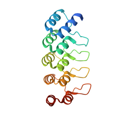
5AAO - PubMed Abstract:
Fluorescent probes constitute a valuable toolbox to address a variety of biological questions and they have become irreplaceable for imaging methods. Commonly, such probes consist of fluorescent proteins or small organic fluorophores coupled to biological molecules of interest. Recently, a novel class of fluorescence-based probes, fluorogen-activating proteins (FAPs), has been reported. These binding proteins are based on antibody single-chain variable fragments and activate fluorogenic dyes, which only become fluorescent upon activation and do not fluoresce when free in solution. Here we present a novel class of fluorogen activators, termed FADAs, based on the very robust designed ankyrin repeat protein scaffold, which also readily folds in the reducing environment of the cytoplasm. The FADA generated in this study was obtained by combined selections with ribosome display and yeast surface display. It enhances the fluorescence of malachite green (MG) dyes by a factor of more than 11,000 and thus activates MG to a similar extent as FAPs based on single-chain variable fragments. As shown by structure determination and in vitro measurements, this FADA was evolved to form a homodimer for the activation of MG dyes. Exploiting the favorable properties of the designed ankyrin repeat protein scaffold, we created a FADA biosensor suitable for imaging of proteins on the cell surface, as well as in the cytosol. Moreover, based on the requirement of dimerization for strong fluorogen activation, a prototype FADA biosensor for in situ detection of a target protein and protein-protein interactions was developed. Therefore, FADAs are versatile fluorescent probes that are easily produced and suitable for diverse applications and thus extend the FAP technology.
Organizational Affiliation:
Department of Biochemistry, University of Zurich, CH-8057, Zurich, Switzerland.