5YXW

Crystal structure of the prefusion form of measles virus fusion protein


Experimental Data Snapshot

  • Method: X-RAY DIFFRACTION
  • Resolution: 2.78 Å
  • R-Value Free: 0.249 
  • R-Value Work: 0.202 
  • R-Value Observed: 0.206 

wwPDB Validation   3D Report Full Report


Ligand Structure Quality Assessment 


This is version 2.1 of the entry. See complete history


Literature

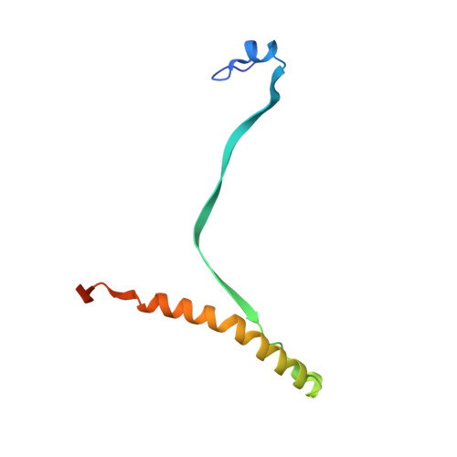
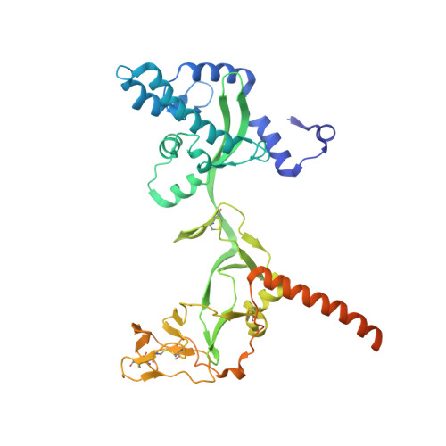
Structures of the prefusion form of measles virus fusion protein in complex with inhibitors.

Hashiguchi, T.Fukuda, Y.Matsuoka, R.Kuroda, D.Kubota, M.Shirogane, Y.Watanabe, S.Tsumoto, K.Kohda, D.Plemper, R.K.Yanagi, Y.

(2018) Proc Natl Acad Sci U S A 115: 2496-2501

  • DOI: https://doi.org/10.1073/pnas.1718957115
  • Primary Citation of Related Structures:  
    5YXW, 5YZC, 5YZD

  • PubMed Abstract: 

    Measles virus (MeV), a major cause of childhood morbidity and mortality, is highly immunotropic and one of the most contagious pathogens. MeV may establish, albeit rarely, persistent infection in the central nervous system, causing fatal and intractable neurodegenerative diseases such as subacute sclerosing panencephalitis and measles inclusion body encephalitis. Recent studies have suggested that particular substitutions in the MeV fusion (F) protein are involved in the pathogenesis by destabilizing the F protein and endowing it with hyperfusogenicity. Here we show the crystal structures of the prefusion MeV-F alone and in complex with the small compound AS-48 or a fusion inhibitor peptide. Notably, these independently developed inhibitors bind the same hydrophobic pocket located at the region connecting the head and stalk of MeV-F, where a number of substitutions in MeV isolates from neurodegenerative diseases are also localized. Since these inhibitors could suppress membrane fusion mediated by most of the hyperfusogenic MeV-F mutants, the development of more effective inhibitors based on the structures may be warranted to treat MeV-induced neurodegenerative diseases.


  • Organizational Affiliation

    Department of Virology, Faculty of Medicine, Kyushu University, 812-8582 Fukuoka, Japan; takaoh@virology.med.kyushu-u.ac.jp yyanagi@virology.med.kyushu-u.ac.jp.


Macromolecules
Find similar proteins by:  (by identity cutoff)  |  3D Structure
Entity ID: 1
MoleculeChains Sequence LengthOrganismDetailsImage
glycoprotein F294Measles virus strain Ichinose-B95aMutation(s): 0 
UniProt
Find proteins for Q786F3 (Measles virus (strain Ichinose-B95a))
Explore Q786F3 
Go to UniProtKB:  Q786F3
Entity Groups  
Sequence Clusters30% Identity50% Identity70% Identity90% Identity95% Identity100% Identity
UniProt GroupQ786F3
Sequence Annotations
Expand
  • Reference Sequence
Find similar proteins by:  (by identity cutoff)  |  3D Structure
Entity ID: 2
MoleculeChains Sequence LengthOrganismDetailsImage
glycoprotein F1,measles virus fusion protein419Measles virus strain Ichinose-B95aMeasles morbillivirus
This entity is chimeric
Mutation(s): 0 
UniProt
Find proteins for Q786F3 (Measles virus (strain Ichinose-B95a))
Explore Q786F3 
Go to UniProtKB:  Q786F3
Entity Groups  
Sequence Clusters30% Identity50% Identity70% Identity90% Identity95% Identity100% Identity
UniProt GroupQ786F3
Sequence Annotations
Expand
  • Reference Sequence
Oligosaccharides

Help

Entity ID: 3
MoleculeChains Length2D Diagram Glycosylation3D Interactions
2-acetamido-2-deoxy-beta-D-glucopyranose-(1-4)-2-acetamido-2-deoxy-beta-D-glucopyranose
C
2N-Glycosylation
Glycosylation Resources
GlyTouCan:  G42666HT
GlyCosmos:  G42666HT
GlyGen:  G42666HT
Small Molecules
Ligands 1 Unique
IDChains Name / Formula / InChI Key2D Diagram3D Interactions
NAG
Query on NAG

Download Ideal Coordinates CCD File 
D [auth A]2-acetamido-2-deoxy-beta-D-glucopyranose
C8 H15 N O6
OVRNDRQMDRJTHS-FMDGEEDCSA-N
Experimental Data & Validation

Experimental Data

  • Method: X-RAY DIFFRACTION
  • Resolution: 2.78 Å
  • R-Value Free: 0.249 
  • R-Value Work: 0.202 
  • R-Value Observed: 0.206 
  • Space Group: I 21 3
Unit Cell:
Length ( Å )Angle ( ˚ )
a = 167.389α = 90
b = 167.389β = 90
c = 167.389γ = 90
Software Package:
Software NamePurpose
PHENIXrefinement
XDSdata reduction
Aimlessdata scaling
PHENIXphasing
Cootmodel building

Structure Validation

View Full Validation Report


Ligand Structure Quality Assessment 


Entry History & Funding Information

Deposition Data


Funding OrganizationLocationGrant Number
MEXTJapan17K19562
MEXTJapan24115005
AMEDJapan17fm0208022h0001
National Institutes of Health/National Institute Of Allergy and Infectious Diseases (NIH/NIAID)United StatesAI071002
National Institutes of Health/National Institute Of Allergy and Infectious Diseases (NIH/NIAID)United StatesHD079327

Revision History  (Full details and data files)

  • Version 1.0: 2018-02-21
    Type: Initial release
  • Version 1.1: 2018-03-07
    Changes: Database references
  • Version 1.2: 2018-03-21
    Changes: Database references
  • Version 2.0: 2020-07-29
    Type: Remediation
    Reason: Carbohydrate remediation
    Changes: Atomic model, Data collection, Derived calculations, Structure summary
  • Version 2.1: 2022-03-23
    Changes: Author supporting evidence, Database references, Structure summary