Three-dimensional structures of bacteriophage neck subunits are shared in Podoviridae, Siphoviridae and Myoviridae
Iwasaki, T., Yamashita, E., Nakagawa, A., Enomoto, A., Tomihara, M., Takeda, S.(2018) Genes Cells 23: 528-536
- PubMed: 29767456
- DOI: https://doi.org/10.1111/gtc.12594
- Primary Citation of Related Structures:
5YDN - PubMed Abstract:
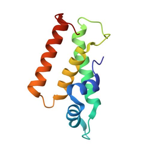
Tailed bacteriophages (Caudovirales) are divided into three families: Myoviridae with long contractile tails, Siphoviridae with long noncontractile tails and Podoviridae with short noncontractile tails. All have an icosahedral head with a portal vertex connected to a neck structure followed by a tail. Bacteriophage Mu belongs to the Myoviridae family. Herein, the gp29 portal subunit and neck subunits gp35, gp36 and gp37 of the Mu phage were purified to elucidate their arrangement in the neck. Both gp29 and gp36 were monomeric in solution, like the corresponding subunits of Podoviridae P22 and Siphoviridae SPP1. X-ray crystal structure of gp36 showed structural similarity to neck subunits of Siphoviridae and Podoviridae. The gp36 structure has a characteristic aromatic hydrophobic core, and the structure of the ring form of the Mu phage connector deduced from the Siphoviridae and Podoviridae connector showed that this feature builds the contact surface between gp36 subunits. Structural comparison with the neck of Siphoviridae and Podoviridae also implies direct interaction between gp36 and gp29. Because gp35 and gp36 form a stable complex, we predict that the head-portal ring (gp29), the connector complex (gp36 and gp35), the tail terminator (gp37) and the tube (gp40) are arranged in the Mu phage neck in this order.
Organizational Affiliation:
Faculty of Science and Technology, Division of Molecular Science, Gunma University, Kiryu, Gunma, Japan.