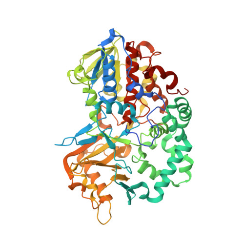
5YB6

L-Amino acid oxidase/monooxygenase from Pseudomonas sp. AIU 813 - L-lysine complex


Experimental Data Snapshot

  • Method: X-RAY DIFFRACTION
  • Resolution: 2.10 Å
  • R-Value Free: 0.283 
  • R-Value Work: 0.234 
  • R-Value Observed: 0.237 

wwPDB Validation   3D Report Full Report


Ligand Structure Quality Assessment 


This is version 1.2 of the entry. See complete history


Literature

Ligand complex structures of l-amino acid oxidase/monooxygenase from

Im, D.Matsui, D.Arakawa, T.Isobe, K.Asano, Y.Fushinobu, S.

(2018) FEBS Open Bio 8: 314-324

  • DOI: https://doi.org/10.1002/2211-5463.12387
  • Primary Citation of Related Structures:  
    5YB6, 5YB7, 5YB8

  • PubMed Abstract: 

    l-Amino acid oxidase/monooxygenase from Pseudomonas sp. AIU 813 (l-AAO/MOG) catalyzes both the oxidative deamination and oxidative decarboxylation of the α-group of l-Lys to produce a keto acid and amide, respectively. l-AAO/MOG exhibits limited specificity for l-amino acid substrates with a basic side chain. We previously determined its ligand-free crystal structure and identified a key residue for maintaining the dual activities. Here, we determined the structures of l-AAO/MOG complexed with l-Lys, l-ornithine, and l-Arg and revealed its substrate recognition. Asp238 is located at the ceiling of a long hydrophobic pocket and forms a strong interaction with the terminal, positively charged group of the substrates. A mutational analysis on the D238A mutant indicated that the interaction is critical for substrate binding but not for catalytic control between the oxidase/monooxygenase activities. The catalytic activities of the D238E mutant unexpectedly increased, while the D238F mutant exhibited altered substrate specificity to long hydrophobic substrates. In the ligand-free structure, there are two channels connecting the active site and solvent, and a short region located at the dimer interface is disordered. In the l-Lys complex structure, a loop region is displaced to plug the channels. Moreover, the disordered region in the ligand-free structure forms a short helix in the substrate complex structures and creates the second binding site for the substrate. It is assumed that the amino acid substrate enters the active site of l-AAO/MOG through this route.


  • Organizational Affiliation

    Department of Biotechnology The University of Tokyo Japan.


Macromolecules
Find similar proteins by:  (by identity cutoff)  |  3D Structure
Entity ID: 1
MoleculeChains Sequence LengthOrganismDetailsImage
L-amino acid oxidase/monooxygenase
A, B, C, D
580Pseudomonas sp. AIU 813Mutation(s): 0 
Gene Names: laaomog
UniProt
Find proteins for W6JQJ6 (Pseudomonas sp. AIU 813)
Explore W6JQJ6 
Go to UniProtKB:  W6JQJ6
Entity Groups  
Sequence Clusters30% Identity50% Identity70% Identity90% Identity95% Identity100% Identity
UniProt GroupW6JQJ6
Sequence Annotations
Expand
  • Reference Sequence
Small Molecules
Ligands 3 Unique
IDChains Name / Formula / InChI Key2D Diagram3D Interactions
FAD
Query on FAD

Download Ideal Coordinates CCD File 
E [auth A],
I [auth B],
M [auth C],
Q [auth D]
FLAVIN-ADENINE DINUCLEOTIDE
C27 H33 N9 O15 P2
VWWQXMAJTJZDQX-UYBVJOGSSA-N
PG6
Query on PG6

Download Ideal Coordinates CCD File 
F [auth A],
J [auth B],
N [auth C],
R [auth D]
1-(2-METHOXY-ETHOXY)-2-{2-[2-(2-METHOXY-ETHOXY]-ETHOXY}-ETHANE
C12 H26 O6
DMDPGPKXQDIQQG-UHFFFAOYSA-N
LYS
Query on LYS

Download Ideal Coordinates CCD File 
G [auth A]
H [auth A]
K [auth B]
L [auth B]
O [auth C]
G [auth A],
H [auth A],
K [auth B],
L [auth B],
O [auth C],
P [auth C],
S [auth D],
T [auth D]
LYSINE
C6 H15 N2 O2
KDXKERNSBIXSRK-YFKPBYRVSA-O
Experimental Data & Validation

Experimental Data

  • Method: X-RAY DIFFRACTION
  • Resolution: 2.10 Å
  • R-Value Free: 0.283 
  • R-Value Work: 0.234 
  • R-Value Observed: 0.237 
  • Space Group: P 1 21 1
Unit Cell:
Length ( Å )Angle ( ˚ )
a = 97.071α = 90
b = 132.293β = 108.67
c = 100.963γ = 90
Software Package:
Software NamePurpose
HKL-2000data processing
HKL-2000data scaling
MOLREPphasing
REFMACrefinement
PDB_EXTRACTdata extraction

Structure Validation

View Full Validation Report


Ligand Structure Quality Assessment 


Entry History 

Deposition Data

Revision History  (Full details and data files)

  • Version 1.0: 2018-02-07
    Type: Initial release
  • Version 1.1: 2018-03-21
    Changes: Database references
  • Version 1.2: 2023-11-22
    Changes: Data collection, Database references, Refinement description