Three Conserved Regions in Baculovirus Sulfhydryl Oxidase P33 Are Critical for Enzymatic Activity and Function
Kuang, W., Zhang, H., Wang, M., Zhou, N.Y., Deng, F., Wang, H., Gong, P., Hu, Z.(2017) J Virol 91
- PubMed: 28904203
- DOI: https://doi.org/10.1128/JVI.01158-17
- Primary Citation of Related Structures:
5XKI, 5XTN, 5XTO, 5XTP, 5XTQ, 5XTR - PubMed Abstract:
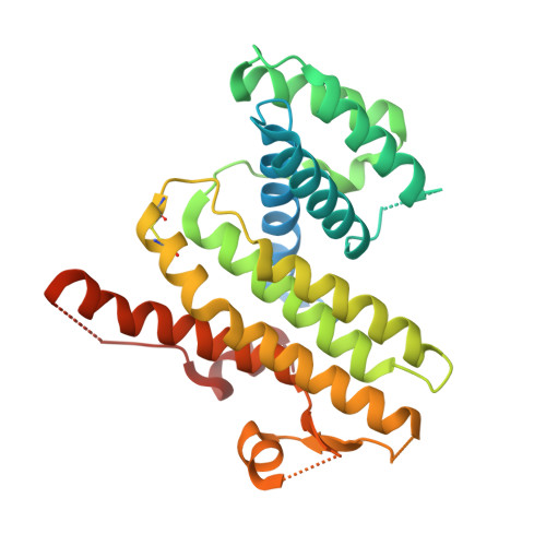
Baculoviruses encode a conserved sulfhydryl oxidase, P33, which is necessary for budded virus (BV) production and multinucleocapsid occlusion-derived virus (ODV) formation. Here, the structural and functional relationship of P33 was revealed by X-ray crystallography, site-directed mutagenesis, and functional analysis. Based on crystallographic characterization and structural analysis, a series of P33 mutants within three conserved regions, i.e., the active site, the dimer interface, and the R127-E183 salt bridge, were constructed. In vitro experiments showed that mutations within the active site and dimer interface severely impaired the sulfhydryl oxidase activity of P33, while the mutations in the salt bridge had a relatively minor influence. Recombinant viruses containing mutated P33 were constructed and assayed in vivo Except for the active-site mutant AXXA, all other mutants produced infectious BVs, although certain mutants had a decreased BV production. The active-site mutant H114A, the dimer interface mutant H227D, and the salt bridge mutant R127A-E183A were further analyzed by electron microscopy and bioassays. The occlusion bodies (OBs) of mutants H114A and R127A-E183A had a ragged surface and contained mostly ODVs with a single nucleocapsid. The OBs of all three mutants contained lower numbers of ODVs and had a significantly reduced oral infectivity in comparison to control virus. Crystallographic analyses further revealed that all three regions may coordinate with one another to achieve optimal function of P33. Taken together, our data revealed that all the three conserved regions are involved in P33 activity and are crucial for virus morphogenesis and peroral infectivity. IMPORTANCE Sulfhydryl oxidase catalyzes disulfide bond formation of substrate proteins. P33, a baculovirus-encoded sulfhydryl oxidase, is different from other cellular and viral sulfhydryl oxidases, bearing unique features in tertiary and quaternary structure organizations. In this study, we found that three conserved regions, i.e., the active site, dimer interface, and the R127-E183 salt bridge, play important roles in the enzymatic activity and function of P33. Previous observations showed that deletion of p33 results in a total loss of budded virus (BV) production and in morphological changes in occlusion-derived virus (ODV). Our study revealed that certain P33 mutants lead to occlusion bodies (OBs) with a ragged surface, decreased embedded ODVs, and reduced oral infectivity. Interestingly, some P33 mutants with impaired ODV/OB still retained BV productivity, indicating that the impacts on BV and on ODV/OB are two distinctly different functions of P33, which are likely to be performed via different substrate proteins.
Organizational Affiliation:
State Key Laboratory of Virology, Wuhan Institute of Virology, Chinese Academy of Sciences, Wuhan, People's Republic of China.