Crystal Structure and Thermostability Characterization of Enterovirus D68 3Dpol
Wang, C., Wang, C., Li, Q., Wang, Z., Xie, W.(2017) J Virol 91
- PubMed: 28659472
- DOI: https://doi.org/10.1128/JVI.00876-17
- Primary Citation of Related Structures:
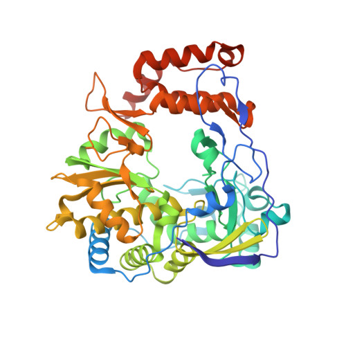
5XE0 - PubMed Abstract:
Enterovirus D68 (EV-D68) is one of the many nonpolio enteroviruses that cause mild to severe respiratory illness. The nonstructural protein 3D pol is an RNA-dependent RNA polymerase (RdRP) of EV-D68 which plays a critical role in the replication of the viral genome and represents a promising drug target. Here, we report the first three-dimensional crystal structure of the RdRP from EV-D68 in complex with the substrate GTP to 2.3-Å resolution. The RdRP structure is similar to structures of other viral RdRPs, where the three domains, termed the palm, fingers, and thumb, form a structure resembling a cupped right hand. Particularly, an N-terminal fragment (Gly1 to Phe30) bridges the fingers and the thumb domains, which accounts for the enhanced stability of the full-length enzyme over the truncation mutant, as assessed by our thermal shift assays and the dynamic light scattering studies. Additionally, the GTP molecule bound proximal to the active site interacts with both the palm and fingers domains to stabilize the core structure of 3D pol Interestingly, using limited proteolysis assays, we found that different nucleoside triphosphates (NTPs) stabilize the polymerase structure by various degrees, with GTP and CTP being the most and least stabilizing nucleosides, respectively. Lastly, we derived a model of the core structure of 3D pol stabilized by GTP, according to our proteolytic studies. The biochemical and biophysical characterizations conducted in this study help us to understand the stability of EV-D68-3D pol , which may extend to other RdRPs as well. IMPORTANCE Enterovirus D68 (EV-D68) is an emerging viral pathogen, which caused sporadic infections around the world. In recent years, epidemiology studies have reported an increasing number of patients with respiratory diseases globally due to the EV-D68 infection. Moreover, the infection has been associated with acute flaccid paralysis and cranial nerve dysfunction in children. However, there are no vaccines and antiviral treatments specifically targeting the virus to date. In this study, we solved the crystal structure of the RNA-dependent RNA polymerase of EV-D68 and carried out systematic biophysical and biochemical characterizations on the overall and local structural stability of the wild-type (WT) enzyme and several variants, which yields a clear view on the structure-activity relationship of the EV-D68 RNA polymerase.
Organizational Affiliation:
School of Pharmaceutical Sciences, The Sun Yat-Sen University, Guangzhou, Guangdong, People's Republic of China.