5X9H

Crystal structure of the Mg2+ channel MgtE in complex with ATP


Experimental Data Snapshot

  • Method: X-RAY DIFFRACTION
  • Resolution: 3.60 Å
  • R-Value Free: 0.283 
  • R-Value Work: 0.250 
  • R-Value Observed: 0.253 

wwPDB Validation   3D Report Full Report


Ligand Structure Quality Assessment 


This is version 1.2 of the entry. See complete history


Literature

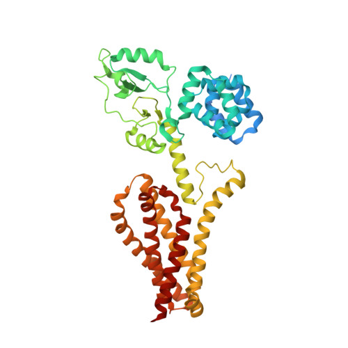
ATP-dependent modulation of MgtE in Mg(2+) homeostasis

Tomita, A.Zhang, M.Jin, F.Zhuang, W.Takeda, H.Maruyama, T.Osawa, M.Hashimoto, K.I.Kawasaki, H.Ito, K.Dohmae, N.Ishitani, R.Shimada, I.Yan, Z.Hattori, M.Nureki, O.

(2017) Nat Commun 8: 148-148

  • DOI: https://doi.org/10.1038/s41467-017-00082-w
  • Primary Citation of Related Structures:  
    5X9G, 5X9H

  • PubMed Abstract: 

    Magnesium is an essential ion for numerous physiological processes. MgtE is a Mg 2+ selective channel involved in the maintenance of intracellular Mg 2+ homeostasis, whose gating is regulated by intracellular Mg 2+ levels. Here, we report that ATP binds to MgtE, regulating its Mg 2+ -dependent gating. Crystal structures of MgtE-ATP complex show that ATP binds to the intracellular CBS domain of MgtE. Functional studies support that ATP binding to MgtE enhances the intracellular domain affinity for Mg 2+ within physiological concentrations of this divalent cation, enabling MgtE to function as an in vivo Mg 2+ sensor. ATP dissociation from MgtE upregulates Mg 2+ influx at both high and low intracellular Mg 2+ concentrations. Using site-directed mutagenesis and structure based-electrophysiological and biochemical analyses, we identify key residues and main structural changes involved in the process. This work provides the molecular basis of ATP-dependent modulation of MgtE in Mg 2+ homeostasis.MgtE is an Mg 2+ transporter involved in Mg 2+ homeostasis. Here, the authors report that ATP regulates the Mg +2 -dependent gating of MgtE and use X-ray crystallography combined with functional studies to propose the molecular mechanisms involved in this process.


  • Organizational Affiliation

    Department of Biological Sciences, Graduate School of Science, The University of Tokyo, 2-11-16 Yayoi, Bunkyo-ku, Tokyo, 113-0032, Japan.


Macromolecules
Find similar proteins by:  (by identity cutoff)  |  3D Structure
Entity ID: 1
MoleculeChains Sequence LengthOrganismDetailsImage
Magnesium transporter MgtE
A, B
473Thermus thermophilus HB8Mutation(s): 0 
Gene Names: TTHA1060
Membrane Entity: Yes 
UniProt
Find proteins for Q5SMG8 (Thermus thermophilus (strain ATCC 27634 / DSM 579 / HB8))
Explore Q5SMG8 
Go to UniProtKB:  Q5SMG8
Entity Groups  
Sequence Clusters30% Identity50% Identity70% Identity90% Identity95% Identity100% Identity
UniProt GroupQ5SMG8
Sequence Annotations
Expand
  • Reference Sequence
Small Molecules
Ligands 2 Unique
IDChains Name / Formula / InChI Key2D Diagram3D Interactions
ATP
Query on ATP

Download Ideal Coordinates CCD File 
C [auth A],
L [auth B]
ADENOSINE-5'-TRIPHOSPHATE
C10 H16 N5 O13 P3
ZKHQWZAMYRWXGA-KQYNXXCUSA-N
MG
Query on MG

Download Ideal Coordinates CCD File 
D [auth A]
E [auth A]
F [auth A]
G [auth A]
H [auth A]
D [auth A],
E [auth A],
F [auth A],
G [auth A],
H [auth A],
I [auth A],
J [auth A],
K [auth A],
M [auth B],
N [auth B],
O [auth B],
P [auth B],
Q [auth B]
MAGNESIUM ION
Mg
JLVVSXFLKOJNIY-UHFFFAOYSA-N
Experimental Data & Validation

Experimental Data

  • Method: X-RAY DIFFRACTION
  • Resolution: 3.60 Å
  • R-Value Free: 0.283 
  • R-Value Work: 0.250 
  • R-Value Observed: 0.253 
  • Space Group: C 1 2 1
Unit Cell:
Length ( Å )Angle ( ˚ )
a = 135.33α = 90
b = 85.651β = 100.02
c = 156.558γ = 90
Software Package:
Software NamePurpose
PHENIXrefinement
XDSdata reduction
XDSdata scaling
MOLREPphasing

Structure Validation

View Full Validation Report


Ligand Structure Quality Assessment 


Entry History & Funding Information

Deposition Data


Funding OrganizationLocationGrant Number
Ministry of Science and Technology of ChinaChina2016YFA0502800

Revision History  (Full details and data files)

  • Version 1.0: 2017-08-16
    Type: Initial release
  • Version 1.1: 2017-08-23
    Changes: Database references
  • Version 1.2: 2024-03-27
    Changes: Data collection, Database references