


Crystal structure of the capping enzyme P5 from Rice Dwarf Virus
Nakamichi, Y., Higashiura, A., Narita, H., Hagiwara, K., Uehara-Ichiki, T., Omura, T., Nakagawa, A.To be published.
Experimental Data Snapshot
Entity ID: 1 | |||||
|---|---|---|---|---|---|
| Molecule | Chains | Sequence Length | Organism | Details | Image |
| mRNA capping enzyme P5 | 804 | Rice dwarf virus (isolate O) | Mutation(s): 0 EC: 2.7.7.50 | 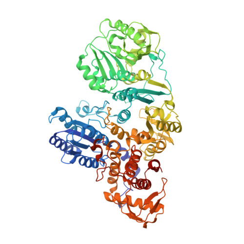 | |
UniProt | |||||
Find proteins for P14583 (Rice dwarf virus (isolate Akita)) Explore P14583 Go to UniProtKB: P14583 | |||||
Entity Groups | |||||
| Sequence Clusters | 30% Identity50% Identity70% Identity90% Identity95% Identity100% Identity | ||||
| UniProt Group | P14583 | ||||
Sequence AnnotationsExpand | |||||
| |||||
| Ligands 1 Unique | |||||
|---|---|---|---|---|---|
| ID | Chains | Name / Formula / InChI Key | 2D Diagram | 3D Interactions | |
| 5GP Query on 5GP | C [auth A], D [auth B] | GUANOSINE-5'-MONOPHOSPHATE C10 H14 N5 O8 P RQFCJASXJCIDSX-UUOKFMHZSA-N |  | ||
| Length ( Å ) | Angle ( ˚ ) |
|---|---|
| a = 76.814 | α = 90 |
| b = 122.631 | β = 90 |
| c = 192.262 | γ = 90 |
| Software Name | Purpose |
|---|---|
| PHENIX | refinement |
| HKL-2000 | data reduction |
| HKL-2000 | data scaling |
| MOLREP | phasing |
| Funding Organization | Location | Grant Number |
|---|---|---|
| JSPS | Japan | JP25251009 |
RCSB PDB (citation) is hosted by
RCSB PDB is a member of the

