Umbrella Sampling and X-ray Crystallographic Analysis Unveil an Arg-Asp Gate Facilitating Inhibitor Binding Inside Phosphopantetheine Adenylyltransferase Allosteric Cleft.
Mondal, A., Chatterjee, R., Datta, S.(2018) J Phys Chem B 122: 1551-1559
- PubMed: 29345931
- DOI: https://doi.org/10.1021/acs.jpcb.7b09543
- Primary Citation of Related Structures:
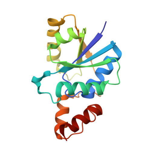
5X6F - PubMed Abstract:
Phosphopantetheine adenylyltransferase (PPAT) is a rate-limiting enzyme essential for biosynthesis of coenzyme A (CoA), which in turn is responsible to regulate the secretion of exotoxins via type III secretion system in Pseudomonas aeruginosa, causing severe health concerns ranging from nosocomial infections to respiratory failure. Acetyl coenzyme A (AcCoA) is a newly reported inhibitor of PPAT, believed to regulate the cellular levels of CoA and thereby the pathogenesis. Very little is known so far regarding the mechanistic details of AcCoA binding inside PPAT-binding cleft. Herein, we have used extensive umbrella sampling simulations to decipher mechanistic insight into the inhibitor accommodation inside the binding cavity. We found that R90 and D94 residues act like a gate near the binding cavity to accommodate and stabilize the incoming ligand. Mutational models concerning these residues also show considerable difference in AcCoA-binding thermodynamics. To substantiate our findings, we have solved the first crystal structure of apo-PPAT from P. aeruginosa, which also found to agree with the simulation results. Collectively, these results describe the mechanistic details of accommodation of inhibitor molecule inside PPAT-binding cavity and also offer valuable insight into regulating cellular levels of CoA/AcCoA and thus controlling the pathogenicity.
Organizational Affiliation:
Structural Biology and Bioinformatics Division, Council of Scientific and Industrial Research-Indian Institute of Chemical Biology , 4 Raja SC Mullick Road, Jadavpur, Kolkata, West Bengal 700032, India.