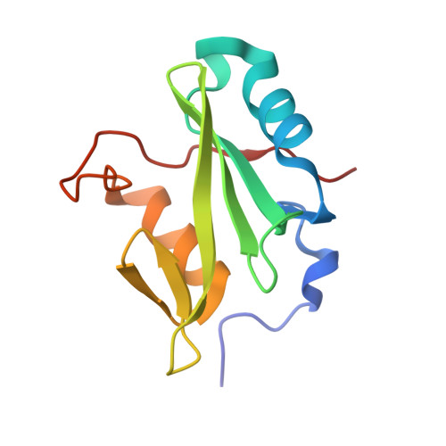
Diversity in peptide recognition by the SH2 domain of SH2B1.
McKercher, M.A., Guan, X., Tan, Z., Wuttke, D.S.(2018) Proteins 86: 164-176
- PubMed: 29127727
- DOI: https://doi.org/10.1002/prot.25420
- Primary Citation of Related Structures:
5W3R - PubMed Abstract:
SH2B1 is a multidomain protein that serves as a key adaptor to regulate numerous cellular events, such as insulin, leptin, and growth hormone signaling pathways. Many of these protein-protein interactions are mediated by the SH2 domain of SH2B1, which recognizes ligands containing a phosphorylated tyrosine (pY), including peptides derived from janus kinase 2, insulin receptor, and insulin receptor substrate-1 and -2. Specificity for the SH2 domain of SH2B1 is conferred in these ligands either by a hydrophobic or an acidic side chain at the +3 position C-terminal to the pY. This specificity for chemically disparate species suggests that SH2B1 relies on distinct thermodynamic or structural mechanisms to bind to peptides. Using binding and structural strategies, we have identified unique thermodynamic signatures for each peptide binding mode, and several SH2B1 residues, including K575 and R578, that play distinct roles in peptide binding. The high-resolution structure of the SH2 domain of SH2B1 further reveals conformationally plastic protein loops that may contribute to the ability of the protein to recognize dissimilar ligands. Together, numerous hydrophobic and electrostatic interactions, in addition to backbone conformational flexibility, permit the recognition of diverse peptides by SH2B1. An understanding of this expanded peptide recognition will allow for the identification of novel physiologically relevant SH2B1/peptide interactions, which can contribute to the design of obesity and diabetes pharmaceuticals to target the ligand-binding interface of SH2B1 with high specificity.
Organizational Affiliation:
Department of Chemistry and Biochemistry, University of Colorado, Boulder, Colorado.