5W2M

APOBEC3F Catalytic Domain Complex with a Single-Stranded DNA


Experimental Data Snapshot

  • Method: X-RAY DIFFRACTION
  • Resolution: 3.70 Å
  • R-Value Free: 0.267 
  • R-Value Work: 0.250 

wwPDB Validation   3D Report Full Report


This is version 1.3 of the entry. See complete history


Literature

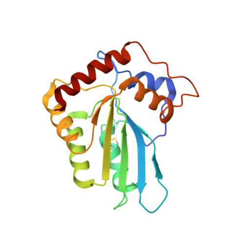
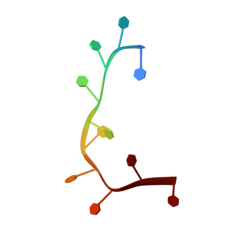
Molecular Interactions of a DNA Modifying Enzyme APOBEC3F Catalytic Domain with a Single-Stranded DNA.

Fang, Y.Xiao, X.Li, S.X.Wolfe, A.Chen, X.S.

(2018) J Mol Biol 430: 87-101

  • DOI: https://doi.org/10.1016/j.jmb.2017.11.007
  • Primary Citation of Related Structures:  
    5W2M

  • PubMed Abstract: 

    The single-stranded DNA (ssDNA) cytidine deaminase APOBEC3F (A3F) deaminates cytosine (C) to uracil (U) and is a known restriction factor of HIV-1. Its C-terminal catalytic domain (CD2) alone is capable of binding single-stranded nucleic acids and is important for deamination. However, little is known about how the CD2 interacts with ssDNA. Here we report a crystal structure of A3F-CD2 in complex with a 10-nucleotide ssDNA composed of poly-thymine, which reveals a novel positively charged nucleic acid binding site distal to the active center that plays a key role in substrate DNA binding and catalytic activity. Lysine and tyrosine residues within this binding site interact with the ssDNA, and mutating these residues dramatically impairs both ssDNA binding and catalytic activity. This binding site is not conserved in APOBEC3G (A3G), which may explain differences in ssDNA-binding characteristics between A3F-CD2 and A3G-CD2. In addition, we observed an alternative Zn-coordination conformation around the active center. These findings reveal the structural relationships between nucleic acid interactions and catalytic activity of A3F.


  • Organizational Affiliation

    Molecular and Computational Biology, Departments of Biological Sciences and Chemistry, University of Southern California, Los Angeles, CA 90089, USA; 161 Hospital of PLA, Wuhan, 430012, China; Department of Clinical Microbiology and Immunology of Southwest Hospital, Third Military Medical University, Chongqing 400038, China.


Macromolecules

Find similar proteins by:  (by identity cutoff)  |  3D Structure
Entity ID: 1
MoleculeChains Sequence LengthOrganismDetailsImage
DNA dC->dU-editing enzyme APOBEC-3F184Homo sapiensMutation(s): 0 
Gene Names: APOBEC3F
EC: 3.5.4
UniProt & NIH Common Fund Data Resources
Find proteins for Q8IUX4 (Homo sapiens)
Explore Q8IUX4 
Go to UniProtKB:  Q8IUX4
PHAROS:  Q8IUX4
GTEx:  ENSG00000128394 
Entity Groups  
Sequence Clusters30% Identity50% Identity70% Identity90% Identity95% Identity100% Identity
UniProt GroupQ8IUX4
Sequence Annotations
Expand
  • Reference Sequence

Find similar nucleic acids by:  Sequence   |   3D Structure  

Entity ID: 2
MoleculeChains LengthOrganismImage
DNA (5'-D(P*TP*TP*TP*TP*TP*TP*TP*TP*TP*T)-3')E,
J [auth N]
10Homo sapiens
Sequence Annotations
Expand
  • Reference Sequence
Experimental Data & Validation

Experimental Data

  • Method: X-RAY DIFFRACTION
  • Resolution: 3.70 Å
  • R-Value Free: 0.267 
  • R-Value Work: 0.250 
  • Space Group: P 1
Unit Cell:
Length ( Å )Angle ( ˚ )
a = 68.194α = 79.6
b = 108.489β = 71.8
c = 108.549γ = 71.75
Software Package:
Software NamePurpose
CNSrefinement
HKL-2000data scaling
PDB_EXTRACTdata extraction
HKL-2000data reduction
MOLREPphasing

Structure Validation

View Full Validation Report


Entry History 

Deposition Data

Revision History  (Full details and data files)

  • Version 1.0: 2017-12-13
    Type: Initial release
  • Version 1.1: 2017-12-20
    Changes: Database references
  • Version 1.2: 2018-01-03
    Changes: Database references
  • Version 1.3: 2023-08-23
    Changes: Data collection, Database references, Structure summary