5UFS

X-Ray Crystal Structure of the Ancestral Glucocorticoid Receptor 2 ligand binding domain in complex with triamcinolone acetonide and SHP coregulator fragment


Experimental Data Snapshot

  • Method: X-RAY DIFFRACTION
  • Resolution: 2.12 Å
  • R-Value Free: 0.250 
  • R-Value Work: 0.215 
  • R-Value Observed: 0.217 

wwPDB Validation   3D Report Full Report


Ligand Structure Quality Assessment 


This is version 1.4 of the entry. See complete history


Literature

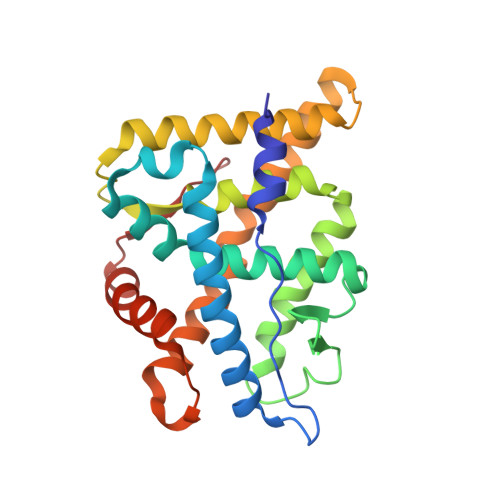
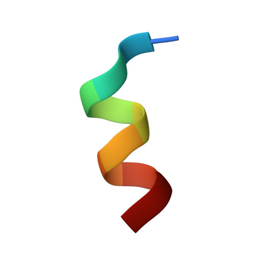
Structural Analysis of the Glucocorticoid Receptor Ligand-Binding Domain in Complex with Triamcinolone Acetonide and a Fragment of the Atypical Coregulator, Small Heterodimer Partner.

Weikum, E.R.Okafor, C.D.D'Agostino, E.H.Colucci, J.K.Ortlund, E.A.

(2017) Mol Pharmacol 92: 12-21

  • DOI: https://doi.org/10.1124/mol.117.108506
  • Primary Citation of Related Structures:  
    5UFS

  • PubMed Abstract: 

    The synthetic glucocorticoids (GCs) dexamethasone, mometasone furoate, and triamcinolone acetonide are pharmaceutical mainstays to treat chronic inflammatory diseases. These drugs bind to the glucocorticoid receptor (GR), a ligand-activated transcription factor and member of the nuclear receptor superfamily. The GR is widely recognized as a therapeutic target for its ability to counter proinflammatory signaling. Despite the popularity of GCs in the clinic, long-term use leads to numerous side effects, driving the need for new and improved drugs with less off-target pharmacology. X-ray crystal structures have played an important role in the drug-design process, permitting the characterization of robust structure-function relationships. However, steroid receptor ligand-binding domains (LBDs) are inherently unstable, and their crystallization requires extensive mutagenesis to enhance expression and crystallization. Here, we use an ancestral variant of GR as a tool to generate a high-resolution crystal structure of GR in complex with the potent glucocorticoid triamcinolone acetonide (TA) and a fragment of the small heterodimer partner (SHP). Using structural analysis, molecular dynamics, and biochemistry, we show that TA increases intramolecular contacts within the LBD to drive affinity and enhance stability of the receptor-ligand complex. These data support the emerging theme that ligand-induced receptor conformational dynamics at the mouth of the pocket play a major role in steroid receptor activation. This work also represents the first GR structure in complex with SHP, which has been suggested to play a role in modulating hepatic GR function.


  • Organizational Affiliation

    Department of Biochemistry, Emory University School of Medicine, Atlanta, Georgia.


Macromolecules
Find similar proteins by:  (by identity cutoff)  |  3D Structure
Entity ID: 1
MoleculeChains Sequence LengthOrganismDetailsImage
Ancestral Glucocorticoid Receptor2
A, B
248unidentifiedMutation(s): 0 
UniProt
Find proteins for A0A1X8XLE9 (unidentified)
Explore A0A1X8XLE9 
Go to UniProtKB:  A0A1X8XLE9
Entity Groups  
Sequence Clusters30% Identity50% Identity70% Identity90% Identity95% Identity100% Identity
UniProt GroupA0A1X8XLE9
Sequence Annotations
Expand
  • Reference Sequence

Find similar proteins by:  Sequence   |   3D Structure  

Entity ID: 2
MoleculeChains Sequence LengthOrganismDetailsImage
SHP NR Box 1 Peptide
C, D
11Homo sapiensMutation(s): 0 
UniProt & NIH Common Fund Data Resources
Find proteins for Q15466 (Homo sapiens)
Explore Q15466 
Go to UniProtKB:  Q15466
PHAROS:  Q15466
GTEx:  ENSG00000131910 
Entity Groups  
Sequence Clusters30% Identity50% Identity70% Identity90% Identity95% Identity100% Identity
UniProt GroupQ15466
Sequence Annotations
Expand
  • Reference Sequence
Small Molecules
Binding Affinity Annotations 
IDSourceBinding Affinity
1TA Binding MOAD:  5UFS Ki: 3.2 (nM) from 1 assay(s)
Experimental Data & Validation

Experimental Data

  • Method: X-RAY DIFFRACTION
  • Resolution: 2.12 Å
  • R-Value Free: 0.250 
  • R-Value Work: 0.215 
  • R-Value Observed: 0.217 
  • Space Group: C 1 2 1
Unit Cell:
Length ( Å )Angle ( ˚ )
a = 87.027α = 90
b = 52.829β = 101.78
c = 126.01γ = 90
Software Package:
Software NamePurpose
HKL-2000data scaling
PHENIXrefinement
PDB_EXTRACTdata extraction
HKL-2000data reduction
PHENIXphasing
DENZOdata reduction
SCALEPACKdata scaling

Structure Validation

View Full Validation Report


Ligand Structure Quality Assessment 


Entry History & Funding Information

Deposition Data


Funding OrganizationLocationGrant Number
National Institutes of Health/National Institute of General Medical Sciences (NIH/NIGMS)United States1F31GM113397-01A1

Revision History  (Full details and data files)

  • Version 1.0: 2017-04-26
    Type: Initial release
  • Version 1.1: 2017-06-07
    Changes: Database references
  • Version 1.2: 2017-09-27
    Changes: Author supporting evidence, Refinement description
  • Version 1.3: 2020-01-01
    Changes: Author supporting evidence
  • Version 1.4: 2023-10-04
    Changes: Data collection, Database references, Refinement description