5TDQ

Crystal Structure of the GOLD domain of ACBD3


Experimental Data Snapshot

  • Method: X-RAY DIFFRACTION
  • Resolution: 2.49 Å
  • R-Value Free: 0.262 
  • R-Value Work: 0.238 
  • R-Value Observed: 0.240 

wwPDB Validation   3D Report Full Report


This is version 1.5 of the entry. See complete history


Literature

The Molecular Basis of Aichi Virus 3A Protein Activation of Phosphatidylinositol 4 Kinase III beta , PI4KB, through ACBD3.

McPhail, J.A.Ottosen, E.H.Jenkins, M.L.Burke, J.E.

(2017) Structure 25: 121-131

  • DOI: https://doi.org/10.1016/j.str.2016.11.016
  • Primary Citation of Related Structures:  
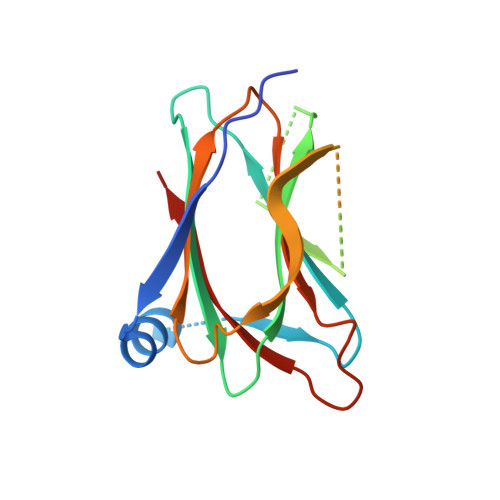
    5TDQ

  • PubMed Abstract: 

    Phosphatidylinositol 4-kinase III beta (PI4KIIIβ) is an essential enzyme in mediating membrane transport, and plays key roles in facilitating viral infection. Many pathogenic positive-sense single-stranded RNA viruses activate PI4KIIIβ to generate phosphatidylinositol 4-phosphate (PI4P)-enriched organelles for viral replication. The molecular basis for PI4KIIIβ activation during viral infection has remained largely unclear. We describe the biochemical reconstitution and characterization of the complex of PI4KIIIβ with the Golgi protein Acyl-coenzyme A binding domain containing protein 3 (ACBD3) and Aichi virus 3A protein on membranes. We find that 3A directly activates PI4KIIIβ, and this activation is sensitized by ACBD3. The interfaces between PI4KIIIβ-ACBD3 and ACBD3-3A were mapped with hydrogen-deuterium exchange mass spectrometry (HDX-MS). Determination of the crystal structure of the ACBD3 GOLD domain revealed a unique N terminus that mediates the interaction with 3A. Rationally designed complex-disrupting mutations in both ACBD3 and PI4KIIIβ completely abrogated the sensitization of 3A activation by ACBD3.


  • Organizational Affiliation

    Department of Biochemistry and Microbiology, University of Victoria, Victoria, BC V8P 5C2, Canada.


Macromolecules
Find similar proteins by:  (by identity cutoff)  |  3D Structure
Entity ID: 1
MoleculeChains Sequence LengthOrganismDetailsImage
Golgi resident protein GCP60166Homo sapiensMutation(s): 0 
Gene Names: ACBD3GCP60GOCAP1GOLPH1
Membrane Entity: Yes 
UniProt & NIH Common Fund Data Resources
Find proteins for Q9H3P7 (Homo sapiens)
Explore Q9H3P7 
Go to UniProtKB:  Q9H3P7
PHAROS:  Q9H3P7
GTEx:  ENSG00000182827 
Entity Groups  
Sequence Clusters30% Identity50% Identity70% Identity90% Identity95% Identity100% Identity
UniProt GroupQ9H3P7
Sequence Annotations
Expand
  • Reference Sequence
Experimental Data & Validation

Experimental Data

  • Method: X-RAY DIFFRACTION
  • Resolution: 2.49 Å
  • R-Value Free: 0.262 
  • R-Value Work: 0.238 
  • R-Value Observed: 0.240 
  • Space Group: P 31 2 1
Unit Cell:
Length ( Å )Angle ( ˚ )
a = 55.51α = 90
b = 55.51β = 90
c = 140.15γ = 120
Software Package:
Software NamePurpose
Aimlessdata scaling
PHASERphasing
PHENIXrefinement
PDB_EXTRACTdata extraction
iMOSFLMdata reduction

Structure Validation

View Full Validation Report


Entry History & Funding Information

Deposition Data


Funding OrganizationLocationGrant Number
Canadian Institutes of Health Research (CIHR)CanadaFRN 142393

Revision History  (Full details and data files)

  • Version 1.0: 2016-11-23
    Type: Initial release
  • Version 1.1: 2017-01-04
    Changes: Database references
  • Version 1.2: 2017-01-18
    Changes: Database references
  • Version 1.3: 2017-09-27
    Changes: Author supporting evidence
  • Version 1.4: 2020-01-08
    Changes: Author supporting evidence
  • Version 1.5: 2023-10-04
    Changes: Data collection, Database references, Refinement description