5TCD

Human alkaline sphingomyelinase (ENPP7) in complex with phosphocholine


Experimental Data Snapshot

  • Method: X-RAY DIFFRACTION
  • Resolution: 2.40 Å
  • R-Value Free: 0.208 
  • R-Value Work: 0.173 
  • R-Value Observed: 0.175 

wwPDB Validation   3D Report Full Report


Ligand Structure Quality Assessment 


This is version 2.0 of the entry. See complete history


Literature

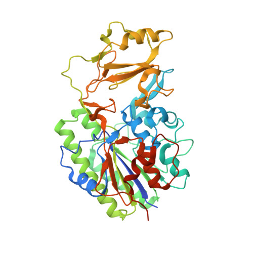
Crystal structure of the human alkaline sphingomyelinase provides insights into substrate recognition.

Gorelik, A.Liu, F.Illes, K.Nagar, B.

(2017) J Biol Chem 292: 7087-7094

  • DOI: https://doi.org/10.1074/jbc.M116.769273
  • Primary Citation of Related Structures:  
    5TCD, 5UDY

  • PubMed Abstract: 

    Absorption of dietary sphingomyelin (SM) requires its initial degradation into ceramide, a process catalyzed by the intestinal enzyme alkaline sphingomyelinase (alk-SMase, NPP7, ENPP7 ). alk-SMase belongs to the nucleotide pyrophosphatase/phosphodiesterase (NPP) family, the members of which hydrolyze nucleoside phosphates, phospholipids, and other related molecules. NPP7 is the only paralog that can cleave SM, and its activity requires the presence of bile salts, a class of physiological anionic detergents. To elucidate the mechanism of substrate recognition, we determined the crystal structure of human alk-SMase in complex with phosphocholine, a reaction product. Although the overall fold and catalytic center are conserved relative to other NPPs, alk-SMase recognizes the choline moiety of its substrates via an NPP7-specific aromatic box composed of tyrosine residues. Mutational analysis and enzymatic activity assays identified features on the surface of the protein-a cationic patch and a unique hydrophobic loop-that are essential for accessing SM in bile salt micelles. These results shed new light on substrate specificity determinants within the NPP enzyme family.


  • Organizational Affiliation

    From the Department of Biochemistry and Groupe de Recherche Axé sur la Structure des Protéines, McGill University, Montreal, Quebec H3G 0B1, Canada.


Macromolecules
Find similar proteins by:  (by identity cutoff)  |  3D Structure
Entity ID: 1
MoleculeChains Sequence LengthOrganismDetailsImage
Ectonucleotide pyrophosphatase/phosphodiesterase family member 7422Homo sapiensMutation(s): 0 
Gene Names: ENPP7UNQ3077/PRO9912
EC: 3.1.4.12
UniProt & NIH Common Fund Data Resources
Find proteins for Q6UWV6 (Homo sapiens)
Explore Q6UWV6 
Go to UniProtKB:  Q6UWV6
PHAROS:  Q6UWV6
GTEx:  ENSG00000182156 
Entity Groups  
Sequence Clusters30% Identity50% Identity70% Identity90% Identity95% Identity100% Identity
UniProt GroupQ6UWV6
Sequence Annotations
Expand
  • Reference Sequence
Oligosaccharides

Help

Entity ID: 2
MoleculeChains Length2D Diagram Glycosylation3D Interactions
2-acetamido-2-deoxy-beta-D-glucopyranose-(1-4)-2-acetamido-2-deoxy-beta-D-glucopyranose
B, C
2N-Glycosylation
Glycosylation Resources
GlyTouCan:  G42666HT
GlyCosmos:  G42666HT
GlyGen:  G42666HT
Entity ID: 3
MoleculeChains Length2D Diagram Glycosylation3D Interactions
beta-D-mannopyranose-(1-4)-2-acetamido-2-deoxy-beta-D-glucopyranose-(1-4)-2-acetamido-2-deoxy-beta-D-glucopyranose
D
3N-Glycosylation
Glycosylation Resources
GlyTouCan:  G15407YE
GlyCosmos:  G15407YE
GlyGen:  G15407YE
Entity ID: 4
MoleculeChains Length2D Diagram Glycosylation3D Interactions
2-acetamido-2-deoxy-beta-D-glucopyranose-(1-4)-[alpha-L-fucopyranose-(1-6)]2-acetamido-2-deoxy-beta-D-glucopyranose
E
3N-Glycosylation
Glycosylation Resources
GlyTouCan:  G21290RB
GlyCosmos:  G21290RB
GlyGen:  G21290RB
Small Molecules
Ligands 5 Unique
IDChains Name / Formula / InChI Key2D Diagram3D Interactions
NAG
Query on NAG

Download Ideal Coordinates CCD File 
H [auth A]2-acetamido-2-deoxy-beta-D-glucopyranose
C8 H15 N O6
OVRNDRQMDRJTHS-FMDGEEDCSA-N
PC
Query on PC

Download Ideal Coordinates CCD File 
UA [auth A]PHOSPHOCHOLINE
C5 H15 N O4 P
YHHSONZFOIEMCP-UHFFFAOYSA-O
IOD
Query on IOD

Download Ideal Coordinates CCD File 
AA [auth A]
BA [auth A]
CA [auth A]
DA [auth A]
EA [auth A]
AA [auth A],
BA [auth A],
CA [auth A],
DA [auth A],
EA [auth A],
FA [auth A],
GA [auth A],
HA [auth A],
IA [auth A],
J [auth A],
JA [auth A],
K [auth A],
KA [auth A],
L [auth A],
LA [auth A],
M [auth A],
MA [auth A],
N [auth A],
NA [auth A],
O [auth A],
OA [auth A],
P [auth A],
PA [auth A],
Q [auth A],
QA [auth A],
R [auth A],
RA [auth A],
S [auth A],
SA [auth A],
T [auth A],
TA [auth A],
U [auth A],
V [auth A],
W [auth A],
X [auth A],
Y [auth A],
Z [auth A]
IODIDE ION
I
XMBWDFGMSWQBCA-UHFFFAOYSA-M
ZN
Query on ZN

Download Ideal Coordinates CCD File 
F [auth A],
G [auth A]
ZINC ION
Zn
PTFCDOFLOPIGGS-UHFFFAOYSA-N
NA
Query on NA

Download Ideal Coordinates CCD File 
I [auth A]SODIUM ION
Na
FKNQFGJONOIPTF-UHFFFAOYSA-N
Experimental Data & Validation

Experimental Data

  • Method: X-RAY DIFFRACTION
  • Resolution: 2.40 Å
  • R-Value Free: 0.208 
  • R-Value Work: 0.173 
  • R-Value Observed: 0.175 
  • Space Group: P 31 2 1
Unit Cell:
Length ( Å )Angle ( ˚ )
a = 104.223α = 90
b = 104.223β = 90
c = 113.893γ = 120
Software Package:
Software NamePurpose
PHENIXrefinement
HKL-2000data reduction
HKL-2000data scaling
PHENIXphasing

Structure Validation

View Full Validation Report


Ligand Structure Quality Assessment 


Entry History & Funding Information

Deposition Data


Funding OrganizationLocationGrant Number
Canadian Institutes of Health Research (CIHR)CanadaMOP-133535

Revision History  (Full details and data files)

  • Version 1.0: 2017-03-22
    Type: Initial release
  • Version 1.1: 2017-05-10
    Changes: Database references
  • Version 1.2: 2020-01-08
    Changes: Author supporting evidence, Data collection
  • Version 2.0: 2020-07-29
    Type: Remediation
    Reason: Carbohydrate remediation
    Changes: Atomic model, Data collection, Derived calculations, Structure summary