5SWI

Crystal structure of SpGH92 in complex with mannose


Experimental Data Snapshot

  • Method: X-RAY DIFFRACTION
  • Resolution: 2.15 Å
  • R-Value Free: 0.221 
  • R-Value Work: 0.197 
  • R-Value Observed: 0.198 

wwPDB Validation   3D Report Full Report


Ligand Structure Quality Assessment 


This is version 1.5 of the entry. See complete history


Literature

Molecular Characterization of N-glycan Degradation and Transport in Streptococcus pneumoniae and Its Contribution to Virulence.

Robb, M.Hobbs, J.K.Woodiga, S.A.Shapiro-Ward, S.Suits, M.D.McGregor, N.Brumer, H.Yesilkaya, H.King, S.J.Boraston, A.B.

(2017) PLoS Pathog 13: e1006090-e1006090

  • DOI: https://doi.org/10.1371/journal.ppat.1006090
  • Primary Citation of Related Structures:  
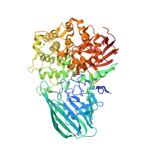
    5SUO, 5SWA, 5SWB, 5SWI

  • PubMed Abstract: 

    The carbohydrate-rich coating of human tissues and cells provide a first point of contact for colonizing and invading bacteria. Commensurate with N-glycosylation being an abundant form of protein glycosylation that has critical functional roles in the host, some host-adapted bacteria possess the machinery to process N-linked glycans. The human pathogen Streptococcus pneumoniae depolymerizes complex N-glycans with enzymes that sequentially trim a complex N-glycan down to the Man3GlcNAc2 core prior to the release of the glycan from the protein by endo-β-N-acetylglucosaminidase (EndoD), which cleaves between the two GlcNAc residues. Here we examine the capacity of S. pneumoniae to process high-mannose N-glycans and transport the products. Through biochemical and structural analyses we demonstrate that S. pneumoniae also possesses an α-(1,2)-mannosidase (SpGH92). This enzyme has the ability to trim the terminal α-(1,2)-linked mannose residues of high-mannose N-glycans to generate Man5GlcNAc2. Through this activity SpGH92 is able to produce a substrate for EndoD, which is not active on high-mannose glycans with α-(1,2)-linked mannose residues. Binding studies and X-ray crystallography show that NgtS, the solute binding protein of an ABC transporter (ABCNG), is able to bind Man5GlcNAc, a product of EndoD activity, with high affinity. Finally, we evaluated the contribution of EndoD and ABCNG to growth of S. pneumoniae on a model N-glycosylated glycoprotein, and the contribution of these enzymes and SpGH92 to virulence in a mouse model. We found that both EndoD and ABCNG contribute to growth of S. pneumoniae, but that only SpGH92 and EndoD contribute to virulence. Therefore, N-glycan processing, but not transport of the released glycan, is required for full virulence in S. pneumoniae. To conclude, we synthesize our findings into a model of N-glycan processing by S. pneumoniae in which both complex and high-mannose N-glycans are targeted, and in which the two arms of this degradation pathway converge at ABCNG.


  • Organizational Affiliation

    Department of Biochemistry and Microbiology, University of Victoria, Victoria, British Columbia, Canada.


Macromolecules
Find similar proteins by:  (by identity cutoff)  |  3D Structure
Entity ID: 1
MoleculeChains Sequence LengthOrganismDetailsImage
Sugar hydrolaseA [auth B],
B [auth A],
C,
D
717Streptococcus pneumoniaeMutation(s): 0 
Gene Names: ERS003549_00384ERS409165_00851ERS409372_00266ERS455085_01031ERS515225_01644
UniProt
Find proteins for A0A0Y0HIE3 (Streptococcus pneumoniae)
Explore A0A0Y0HIE3 
Go to UniProtKB:  A0A0Y0HIE3
Entity Groups  
Sequence Clusters30% Identity50% Identity70% Identity90% Identity95% Identity100% Identity
UniProt GroupA0A0Y0HIE3
Sequence Annotations
Expand
  • Reference Sequence
Small Molecules
Ligands 3 Unique
IDChains Name / Formula / InChI Key2D Diagram3D Interactions
BMA
Query on BMA

Download Ideal Coordinates CCD File 
AA [auth D],
J [auth B],
R [auth A],
V [auth C]
beta-D-mannopyranose
C6 H12 O6
WQZGKKKJIJFFOK-RWOPYEJCSA-N
GOL
Query on GOL

Download Ideal Coordinates CCD File 
F [auth B]
G [auth B]
H [auth B]
I [auth B]
K [auth A]
F [auth B],
G [auth B],
H [auth B],
I [auth B],
K [auth A],
L [auth A],
M [auth A],
N [auth A],
O [auth A],
P [auth A],
Q [auth A],
T [auth C],
U [auth C],
X [auth D],
Y [auth D],
Z [auth D]
GLYCEROL
C3 H8 O3
PEDCQBHIVMGVHV-UHFFFAOYSA-N
CA
Query on CA

Download Ideal Coordinates CCD File 
BA [auth D],
E [auth B],
S [auth A],
W [auth C]
CALCIUM ION
Ca
BHPQYMZQTOCNFJ-UHFFFAOYSA-N
Experimental Data & Validation

Experimental Data

  • Method: X-RAY DIFFRACTION
  • Resolution: 2.15 Å
  • R-Value Free: 0.221 
  • R-Value Work: 0.197 
  • R-Value Observed: 0.198 
  • Space Group: P 21 21 21
Unit Cell:
Length ( Å )Angle ( ˚ )
a = 130.003α = 90
b = 161.711β = 90
c = 208.404γ = 90
Software Package:
Software NamePurpose
REFMACrefinement
SCALAdata scaling
PHASERphasing
PDB_EXTRACTdata extraction
XDSdata reduction

Structure Validation

View Full Validation Report


Ligand Structure Quality Assessment 


Entry History & Funding Information

Deposition Data


Funding OrganizationLocationGrant Number
Canadian Institutes of Health Research (CIHR)CanadaMOP 130305

Revision History  (Full details and data files)

  • Version 1.0: 2016-12-14
    Type: Initial release
  • Version 1.1: 2017-01-18
    Changes: Database references
  • Version 1.2: 2017-09-20
    Changes: Author supporting evidence
  • Version 1.3: 2020-01-08
    Changes: Author supporting evidence
  • Version 1.4: 2020-07-29
    Type: Remediation
    Reason: Carbohydrate remediation
    Changes: Data collection, Derived calculations, Structure summary
  • Version 1.5: 2023-10-04
    Changes: Data collection, Database references, Refinement description, Structure summary