5ODL

Single-stranded DNA-binding protein from bacteriophage Enc34 in complex with ssDNA


Experimental Data Snapshot

  • Method: X-RAY DIFFRACTION
  • Resolution: 1.56 Å
  • R-Value Free: 0.191 
  • R-Value Work: 0.141 
  • R-Value Observed: 0.143 

wwPDB Validation   3D Report Full Report


This is version 1.2 of the entry. See complete history


Literature

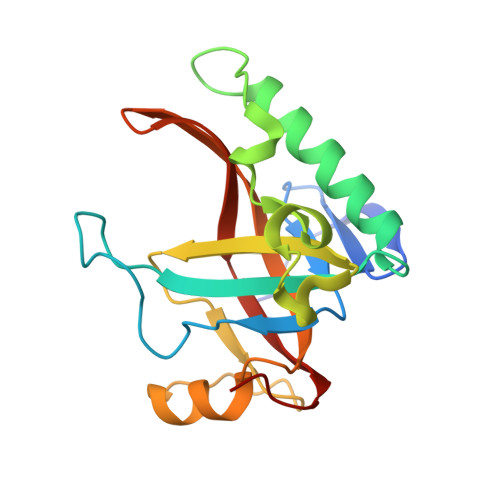
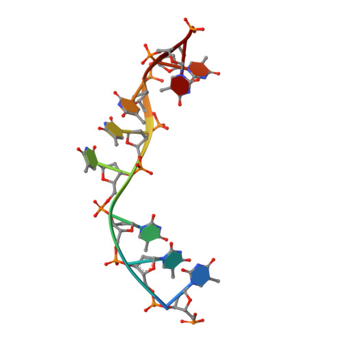
Structural Basis for DNA Recognition of a Single-stranded DNA-binding Protein from Enterobacter Phage Enc34.

Cernooka, E.Rumnieks, J.Tars, K.Kazaks, A.

(2017) Sci Rep 7: 15529-15529

  • DOI: https://doi.org/10.1038/s41598-017-15774-y
  • Primary Citation of Related Structures:  
    5ODJ, 5ODK, 5ODL

  • PubMed Abstract: 

    Modern DNA sequencing capabilities have led to the discovery of a large number of new bacteriophage genomes, which are a rich source of novel proteins with an unidentified biological role. The genome of Enterobacter cancerogenus bacteriophage Enc34 contains several proteins of unknown function that are nevertheless conserved among distantly related phages. Here, we report the crystal structure of a conserved Enc34 replication protein ORF6 which contains a domain of unknown function DUF2815. Despite the low (~15%) sequence identity, the Enc34 ORF6 structurally resembles the gene 2.5 protein from bacteriophage T7, and likewise is a single-stranded DNA (ssDNA)-binding protein (SSB) that consists of a variation of the oligosaccharide/oligonucleotide-binding (OB)-fold and an unstructured C-terminal segment. We further report the crystal structure of a C-terminally truncated ORF6 in complex with an ssDNA oligonucleotide that reveals a DNA-binding mode involving two aromatic stacks and multiple electrostatic interactions, with implications for a common ssDNA recognition mechanism for all T7-type SSBs.


  • Organizational Affiliation

    Latvian Biomedical Research and Study Centre, Riga, LV-1067, Latvia.


Macromolecules

Find similar proteins by:  (by identity cutoff)  |  3D Structure
Entity ID: 1
MoleculeChains Sequence LengthOrganismDetailsImage
single-stranded DNA-binding protein180Hafnia phage Enc34Mutation(s): 0 
UniProt
Find proteins for H6WYG2 (Enterobacter phage Enc34)
Explore H6WYG2 
Go to UniProtKB:  H6WYG2
Entity Groups  
Sequence Clusters30% Identity50% Identity70% Identity90% Identity95% Identity100% Identity
UniProt GroupH6WYG2
Sequence Annotations
Expand
  • Reference Sequence

Find similar nucleic acids by:  Sequence   |   3D Structure  

Entity ID: 2
MoleculeChains LengthOrganismImage
oligo(T)B [auth D]9synthetic construct
Sequence Annotations
Expand
  • Reference Sequence
Experimental Data & Validation

Experimental Data

  • Method: X-RAY DIFFRACTION
  • Resolution: 1.56 Å
  • R-Value Free: 0.191 
  • R-Value Work: 0.141 
  • R-Value Observed: 0.143 
  • Space Group: P 61
Unit Cell:
Length ( Å )Angle ( ˚ )
a = 43.489α = 90
b = 43.489β = 90
c = 179.106γ = 120
Software Package:
Software NamePurpose
REFMACrefinement
iMOSFLMdata reduction
SCALAdata scaling
PHASERphasing

Structure Validation

View Full Validation Report


Entry History 

Deposition Data

Revision History  (Full details and data files)

  • Version 1.0: 2017-10-25
    Type: Initial release
  • Version 1.1: 2017-11-29
    Changes: Database references
  • Version 1.2: 2024-01-17
    Changes: Data collection, Database references, Refinement description