5O2N

Lytic transglycosylase in action


Experimental Data Snapshot

  • Method: X-RAY DIFFRACTION
  • Resolution: 1.51 Å
  • R-Value Free: 0.192 
  • R-Value Work: 0.162 
  • R-Value Observed: 0.163 

wwPDB Validation   3D Report Full Report


Ligand Structure Quality Assessment 


This is version 2.0 of the entry. See complete history

Re-refinement Note

This entry reflects an alternative modeling of the original data in: 4YP4


Literature

A step-by-stepin crystalloguide to bond cleavage and 1,6-anhydro-sugar product synthesis by a peptidoglycan-degrading lytic transglycosylase.

Williams, A.H.Wheeler, R.Rateau, L.Malosse, C.Chamot-Rooke, J.Haouz, A.Taha, M.K.Boneca, I.G.

(2018) J Biol Chem 293: 6000-6010

  • DOI: https://doi.org/10.1074/jbc.RA117.001095
  • Primary Citation of Related Structures:  
    5O1J, 5O24, 5O29, 5O2N, 6FPN

  • PubMed Abstract: 

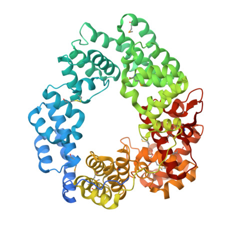
    Lytic transglycosylases (LTs) are a class of enzymes important for the recycling and metabolism of peptidoglycan (PG). LTs cleave the β-1,4-glycosidic bond between N -acetylmuramic acid (MurNAc) and GlcNAc in the PG glycan strand, resulting in the concomitant formation of 1,6-anhydro- N -acetylmuramic acid and GlcNAc. No LTs reported to date have utilized chitins as substrates, despite the fact that chitins are GlcNAc polymers linked via β-1,4-glycosidic bonds, which are the known site of chemical activity for LTs. Here, we demonstrate enzymatically that LtgA, a non-canonical, substrate-permissive LT from Neisseria meningitidis utilizes chitopentaose ((GlcNAc) 5 ) as a substrate to produce three newly identified sugars: 1,6-anhydro-chitobiose, 1,6-anhydro-chitotriose, and 1,6-anhydro-chitotetraose. Although LTs have been widely studied, their complex reactions have not previously been visualized in the crystalline state because macromolecular PG is insoluble. Here, we visualized the cleavage of the glycosidic bond and the liberation of GlcNAc-derived residues by LtgA, followed by the synthesis of atypical 1,6-anhydro-GlcNAc derivatives. In addition to the newly identified anhydro-chitin products, we identified trapped intermediates, unpredicted substrate rearrangements, sugar distortions, and a conserved crystallographic water molecule bound to the catalytic glutamate of a high-resolution native LT. This study enabled us to propose a revised alternative mechanism for LtgA that could also be applicable to other LTs. Our work contributes to the understanding of the mechanisms of LTs in bacterial cell wall biology.


  • Organizational Affiliation

    From the Institut Pasteur, Département de Microbiologie, Unité Biologie et Génétique de la Paroi Bactérienne, 75015 Paris, France, awilliam@pasteur.fr.


Macromolecules
Find similar proteins by:  (by identity cutoff)  |  3D Structure
Entity ID: 1
MoleculeChains Sequence LengthOrganismDetailsImage
Transglycosylase616Neisseria meningitidisMutation(s): 0 
Gene Names: sltERS514729_01258
EC: 4.2.2
UniProt
Find proteins for Q9JXP1 (Neisseria meningitidis serogroup B (strain MC58))
Explore Q9JXP1 
Go to UniProtKB:  Q9JXP1
Entity Groups  
Sequence Clusters30% Identity50% Identity70% Identity90% Identity95% Identity100% Identity
UniProt GroupQ9JXP1
Sequence Annotations
Expand
  • Reference Sequence
Oligosaccharides

Help

Entity ID: 2
MoleculeChains Length2D Diagram Glycosylation3D Interactions
2-acetamido-2-deoxy-beta-D-glucopyranose-(1-4)-2-acetamido-2-deoxy-beta-D-glucopyranose-(1-4)-2-acetamido-2-deoxy-beta-D-glucopyranose-(1-4)-2-acetamido-2-deoxy-alpha-D-glucopyranose
B
4N/A
Glycosylation Resources
GlyTouCan:  G93613RO
GlyCosmos:  G93613RO
GlyGen:  G93613RO
Small Molecules
Ligands 2 Unique
IDChains Name / Formula / InChI Key2D Diagram3D Interactions
NAG
Query on NAG

Download Ideal Coordinates CCD File 
C [auth A]2-acetamido-2-deoxy-beta-D-glucopyranose
C8 H15 N O6
OVRNDRQMDRJTHS-FMDGEEDCSA-N
FLC
Query on FLC

Download Ideal Coordinates CCD File 
D [auth A]CITRATE ANION
C6 H5 O7
KRKNYBCHXYNGOX-UHFFFAOYSA-K
Modified Residues  1 Unique
IDChains TypeFormula2D DiagramParent
MSE
Query on MSE
A
L-PEPTIDE LINKINGC5 H11 N O2 SeMET
Experimental Data & Validation

Experimental Data

  • Method: X-RAY DIFFRACTION
  • Resolution: 1.51 Å
  • R-Value Free: 0.192 
  • R-Value Work: 0.162 
  • R-Value Observed: 0.163 
  • Space Group: P 21 21 21
Unit Cell:
Length ( Å )Angle ( ˚ )
a = 67.1α = 90
b = 72.6β = 90
c = 125γ = 90
Software Package:
Software NamePurpose
PHENIXrefinement
XDSdata reduction
XDSdata scaling
PHENIXphasing

Structure Validation

View Full Validation Report


Ligand Structure Quality Assessment 


Entry History 

Deposition Data

Revision History  (Full details and data files)

  • Version 1.0: 2018-03-14
    Type: Initial release
  • Version 1.1: 2018-05-02
    Changes: Data collection, Database references
  • Version 1.2: 2018-11-28
    Changes: Data collection, Structure summary
  • Version 1.3: 2019-10-16
    Changes: Data collection
  • Version 2.0: 2020-07-29
    Type: Remediation
    Reason: Carbohydrate remediation
    Changes: Atomic model, Data collection, Derived calculations, Structure summary