Structural Study of the C-Terminal Domain of Nonstructural Protein 1 from Japanese Encephalitis Virus.
Poonsiri, T., Wright, G.S.A., Diamond, M.S., Turtle, L., Solomon, T., Antonyuk, S.V.(2018) J Virol 92
- PubMed: 29343583
- DOI: https://doi.org/10.1128/JVI.01868-17
- Primary Citation of Related Structures:
5O19, 5O36 - PubMed Abstract:
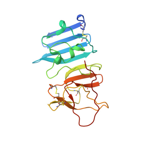
Japanese encephalitis virus (JEV) is a mosquito-transmitted flavivirus that is closely related to other emerging viral pathogens, including dengue virus (DENV), West Nile virus (WNV), and Zika virus (ZIKV). JEV infection can result in meningitis and encephalitis, which in severe cases cause permanent brain damage and death. JEV occurs predominantly in rural areas throughout Southeast Asia, the Pacific Islands, and the Far East, causing around 68,000 cases of infection worldwide each year. In this report, we present a 2.1-Å-resolution crystal structure of the C-terminal β-ladder domain of JEV nonstructural protein 1 (NS1-C). The surface charge distribution of JEV NS1-C is similar to those of WNV and ZIKV but differs from that of DENV. Analysis of the JEV NS1-C structure, with in silico molecular dynamics simulation and experimental solution small-angle X-ray scattering, indicates extensive loop flexibility on the exterior of the protein. This, together with the surface charge distribution, indicates that flexibility influences the protein-protein interactions that govern pathogenicity. These factors also affect the interaction of NS1 with the 22NS1 monoclonal antibody, which is protective against West Nile virus infection. Liposome and heparin binding assays indicate that only the N-terminal region of NS1 mediates interaction with membranes and that sulfate binding sites common to NS1 structures are not glycosaminoglycan binding interfaces. This report highlights several differences between flavivirus NS1 proteins and contributes to our understanding of their structure-pathogenic function relationships. IMPORTANCE JEV is a major cause of viral encephalitis in Asia. Despite extensive vaccination, epidemics still occur. Nonstructural protein 1 (NS1) plays a role in viral replication, and, because it is secreted, it can exhibit a wide range of interactions with host proteins. NS1 sequence and protein folds are conserved within the Flavivirus genus, but variations in NS1 protein-protein interactions among viruses likely contribute to differences in pathogenesis. Here, we compared characteristics of the C-terminal β-ladder domain of NS1 between flaviviruses, including surface charge, loop flexibility, epitope cross-reactivity, membrane adherence, and glycosaminoglycan binding. These structural features are central to NS1 functionality and may provide insight into the development of diagnostic tests and therapeutics.
Organizational Affiliation:
Molecular Biophysics Group, Institute of Integrative Biology, Faculty of Health and Life Sciences, University of Liverpool, Liverpool, United Kingdom.