5N6I

Crystal structure of mouse cGAS in complex with 39 bp DNA


Experimental Data Snapshot

  • Method: X-RAY DIFFRACTION
  • Resolution: 3.60 Å
  • R-Value Free: 0.256 
  • R-Value Work: 0.204 
  • R-Value Observed: 0.206 

wwPDB Validation   3D Report Full Report


This is version 1.5 of the entry. See complete history


Literature

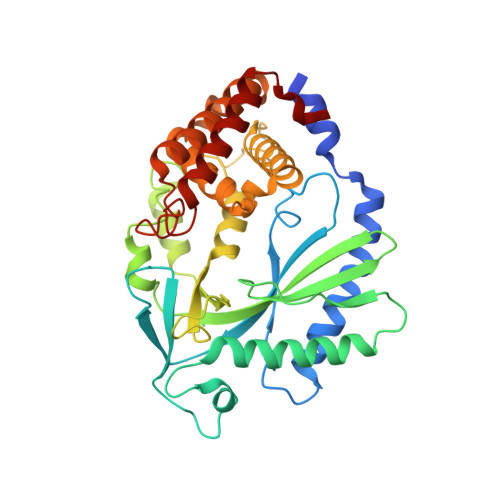
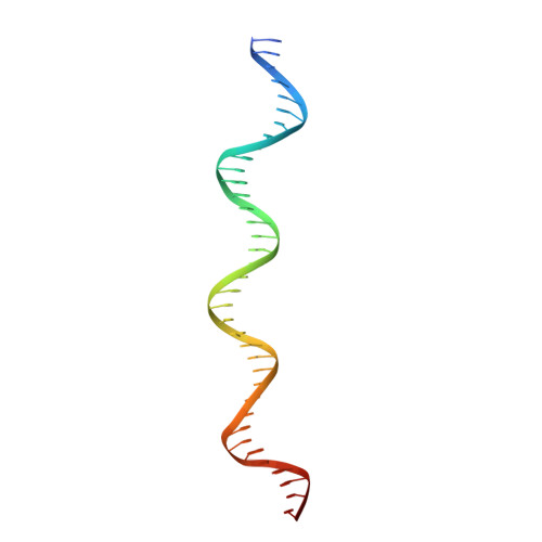
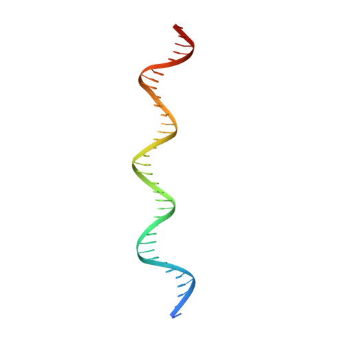
cGAS senses long and HMGB/TFAM-bound U-turn DNA by forming protein-DNA ladders.

Andreeva, L.Hiller, B.Kostrewa, D.Lassig, C.de Oliveira Mann, C.C.Jan Drexler, D.Maiser, A.Gaidt, M.Leonhardt, H.Hornung, V.Hopfner, K.P.

(2017) Nature 549: 394-398

  • DOI: https://doi.org/10.1038/nature23890
  • Primary Citation of Related Structures:  
    5N6I

  • PubMed Abstract: 

    Cytosolic DNA arising from intracellular pathogens triggers a powerful innate immune response. It is sensed by cyclic GMP-AMP synthase (cGAS), which elicits the production of type I interferons by generating the second messenger 2'3'-cyclic-GMP-AMP (cGAMP). Endogenous nuclear or mitochondrial DNA can also be sensed by cGAS under certain conditions, resulting in sterile inflammation. The cGAS dimer binds two DNA ligands shorter than 20 base pairs side-by-side, but 20-base-pair DNA fails to activate cGAS in vivo and is a poor activator in vitro. Here we show that cGAS is activated in a strongly DNA length-dependent manner both in vitro and in human cells. We also show that cGAS dimers form ladder-like networks with DNA, leading to cooperative sensing of DNA length: assembly of the pioneering cGAS dimer between two DNA molecules is ineffective; but, once formed, it prearranges the flanking DNA to promote binding of subsequent cGAS dimers. Remarkably, bacterial and mitochondrial nucleoid proteins HU and mitochondrial transcription factor A (TFAM), as well as high-mobility group box 1 protein (HMGB1), can strongly stimulate long DNA sensing by cGAS. U-turns and bends in DNA induced by these proteins pre-structure DNA to nucleate cGAS dimers. Our results suggest a nucleation-cooperativity-based mechanism for sensitive detection of mitochondrial DNA and pathogen genomes, and identify HMGB/TFAM proteins as DNA-structuring host factors. They provide an explanation for the peculiar cGAS dimer structure and suggest that cGAS preferentially binds incomplete nucleoid-like structures or bent DNA.


  • Organizational Affiliation

    Department of Biochemistry, Ludwig-Maximilians-Universität München, 81377 Munich, Germany.


Macromolecules

Find similar proteins by:  (by identity cutoff)  |  3D Structure
Entity ID: 1
MoleculeChains Sequence LengthOrganismDetailsImage
Cyclic GMP-AMP synthase
A, B, C, D, E
A, B, C, D, E, F
370Mus musculusMutation(s): 0 
Gene Names: Mb21d1
EC: 2.7.7.86
UniProt & NIH Common Fund Data Resources
Find proteins for Q8C6L5 (Mus musculus)
Explore Q8C6L5 
Go to UniProtKB:  Q8C6L5
IMPC:  MGI:2442261
Entity Groups  
Sequence Clusters30% Identity50% Identity70% Identity90% Identity95% Identity100% Identity
UniProt GroupQ8C6L5
Sequence Annotations
Expand
  • Reference Sequence
Find similar nucleic acids by:  (by identity cutoff)  |  3D Structure
Entity ID: 2
MoleculeChains LengthOrganismImage
DNA (37-MER)
G, I, K, M
39synthetic construct
Sequence Annotations
Expand
  • Reference Sequence
Find similar nucleic acids by:  (by identity cutoff)  |  3D Structure
Entity ID: 3
MoleculeChains LengthOrganismImage
DNA (36-MER)
H, J, L, N
39synthetic construct
Sequence Annotations
Expand
  • Reference Sequence
Experimental Data & Validation

Experimental Data

  • Method: X-RAY DIFFRACTION
  • Resolution: 3.60 Å
  • R-Value Free: 0.256 
  • R-Value Work: 0.204 
  • R-Value Observed: 0.206 
  • Space Group: C 1 2 1
Unit Cell:
Length ( Å )Angle ( ˚ )
a = 168.53α = 90
b = 122.94β = 96.37
c = 179.98γ = 90
Software Package:
Software NamePurpose
REFMACrefinement
XDSdata reduction
XSCALEdata scaling
PHASERphasing
STARANISOdata scaling

Structure Validation

View Full Validation Report


Entry History & Funding Information

Deposition Data


Funding OrganizationLocationGrant Number
German Research FoundationGermanyHO2489/8-1
the Center for Integrated Protein SciencesGermany--

Revision History  (Full details and data files)

  • Version 1.0: 2017-09-13
    Type: Initial release
  • Version 1.1: 2017-09-27
    Changes: Database references
  • Version 1.2: 2017-10-04
    Changes: Database references
  • Version 1.3: 2018-01-24
    Changes: Source and taxonomy
  • Version 1.4: 2019-10-16
    Changes: Data collection
  • Version 1.5: 2024-01-17
    Changes: Data collection, Database references, Refinement description