5MJM

Single-shot pink beam serial crystallography: Phycocyanin (Five chips merged)


Experimental Data Snapshot

  • Method: X-RAY DIFFRACTION
  • Resolution: 2.30 Å
  • R-Value Free: 0.181 
  • R-Value Work: 0.139 
  • R-Value Observed: 0.143 

wwPDB Validation   3D Report Full Report


Ligand Structure Quality Assessment 


This is version 1.1 of the entry. See complete history


Literature

Pink-beam serial crystallography.

Meents, A.Wiedorn, M.O.Srajer, V.Henning, R.Sarrou, I.Bergtholdt, J.Barthelmess, M.Reinke, P.Y.A.Dierksmeyer, D.Tolstikova, A.Schaible, S.Messerschmidt, M.Ogata, C.M.Kissick, D.J.Taft, M.H.Manstein, D.J.Lieske, J.Oberthuer, D.Fischetti, R.F.Chapman, H.N.

(2017) Nat Commun 8: 1281-1281

  • DOI: https://doi.org/10.1038/s41467-017-01417-3
  • Primary Citation of Related Structures:  
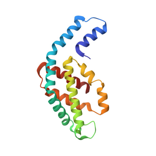
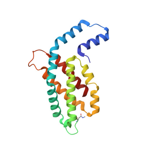
    5MJL, 5MJM, 5MJP, 5MJQ, 5O7M

  • PubMed Abstract: 

    Serial X-ray crystallography allows macromolecular structure determination at both X-ray free electron lasers (XFELs) and, more recently, synchrotron sources. The time resolution for serial synchrotron crystallography experiments has been limited to millisecond timescales with monochromatic beams. The polychromatic, "pink", beam provides a more than two orders of magnitude increased photon flux and hence allows accessing much shorter timescales in diffraction experiments at synchrotron sources. Here we report the structure determination of two different protein samples by merging pink-beam diffraction patterns from many crystals, each collected with a single 100 ps X-ray pulse exposure per crystal using a setup optimized for very low scattering background. In contrast to experiments with monochromatic radiation, data from only 50 crystals were required to obtain complete datasets. The high quality of the diffraction data highlights the potential of this method for studying irreversible reactions at sub-microsecond timescales using high-brightness X-ray facilities.


  • Organizational Affiliation

    Center for Free Electron Laser Science, DESY, Notkestrasse 85, 22607, Hamburg, Germany. alke.meents@desy.de.


Macromolecules
Find similar proteins by:  (by identity cutoff)  |  3D Structure
Entity ID: 1
MoleculeChains Sequence LengthOrganismDetailsImage
C-phycocyanin alpha chain162Thermosynechococcus vestitus BP-1Mutation(s): 0 
UniProt
Find proteins for P50032 (Thermosynechococcus vestitus (strain NIES-2133 / IAM M-273 / BP-1))
Explore P50032 
Go to UniProtKB:  P50032
Entity Groups  
Sequence Clusters30% Identity50% Identity70% Identity90% Identity95% Identity100% Identity
UniProt GroupP50032
Sequence Annotations
Expand
  • Reference Sequence
Find similar proteins by:  (by identity cutoff)  |  3D Structure
Entity ID: 2
MoleculeChains Sequence LengthOrganismDetailsImage
C-phycocyanin beta chain172Thermosynechococcus vestitus BP-1Mutation(s): 0 
UniProt
Find proteins for P50033 (Thermosynechococcus vestitus (strain NIES-2133 / IAM M-273 / BP-1))
Explore P50033 
Go to UniProtKB:  P50033
Entity Groups  
Sequence Clusters30% Identity50% Identity70% Identity90% Identity95% Identity100% Identity
UniProt GroupP50033
Sequence Annotations
Expand
  • Reference Sequence
Small Molecules
Modified Residues  1 Unique
IDChains TypeFormula2D DiagramParent
MEN
Query on MEN
B
L-PEPTIDE LINKINGC5 H10 N2 O3ASN
Experimental Data & Validation

Experimental Data

  • Method: X-RAY DIFFRACTION
  • Resolution: 2.30 Å
  • R-Value Free: 0.181 
  • R-Value Work: 0.139 
  • R-Value Observed: 0.143 
  • Space Group: H 3 2
Unit Cell:
Length ( Å )Angle ( ˚ )
a = 187.8α = 90
b = 187.8β = 90
c = 60.7γ = 120
Software Package:
Software NamePurpose
PHENIXrefinement
PHASERphasing
Epinormdata reduction
Precognitiondata reduction

Structure Validation

View Full Validation Report


Ligand Structure Quality Assessment 


Entry History 

Deposition Data

Revision History  (Full details and data files)

  • Version 1.0: 2017-11-15
    Type: Initial release
  • Version 1.1: 2024-01-17
    Changes: Data collection, Database references, Refinement description