Conformational switch of the bacterial adhesin FimH in the absence of the regulatory domain: Engineering a minimalistic allosteric system.
Rabbani, S., Fiege, B., Eris, D., Silbermann, M., Jakob, R.P., Navarra, G., Maier, T., Ernst, B.(2018) J Biol Chem 293: 1835-1849
- PubMed: 29180452
- DOI: https://doi.org/10.1074/jbc.M117.802942
- Primary Citation of Related Structures:
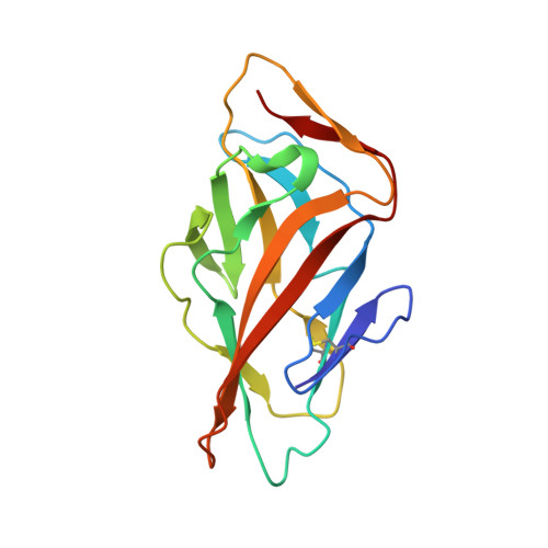
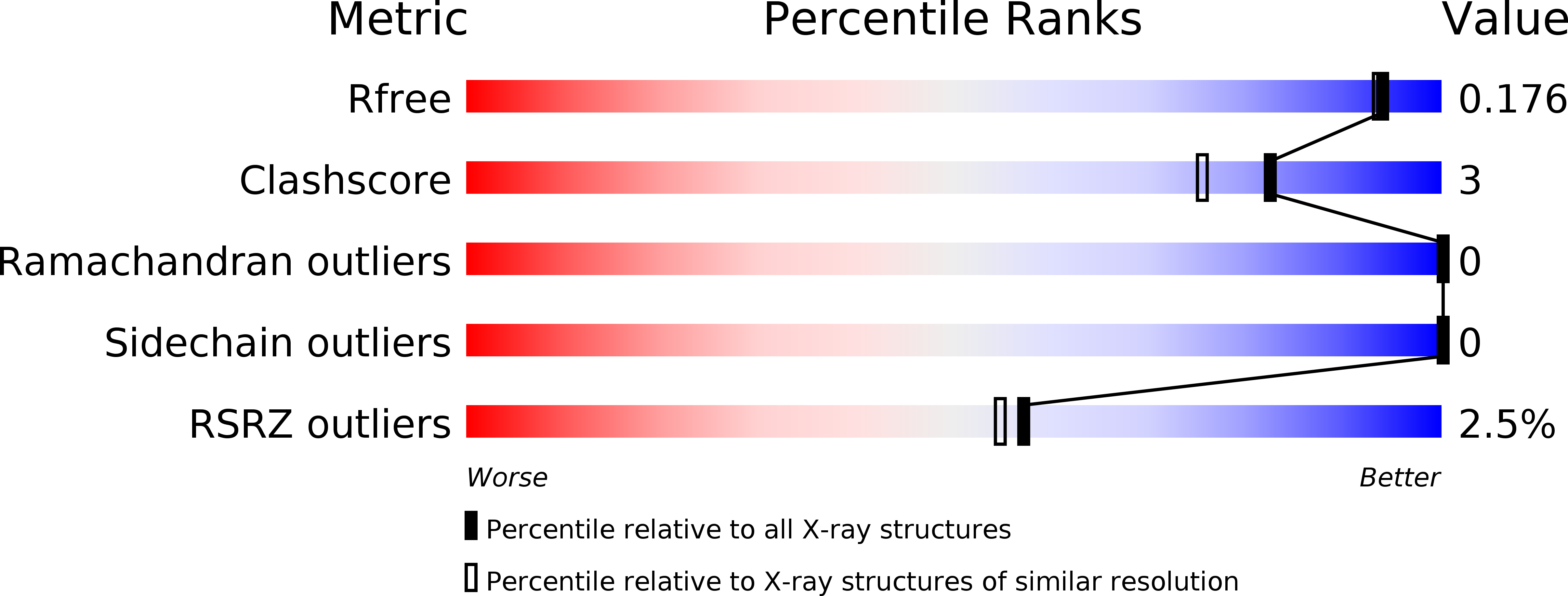
5MCA - PubMed Abstract:
For many biological processes such as ligand binding, enzymatic catalysis, or protein folding, allosteric regulation of protein conformation and dynamics is fundamentally important. One example is the bacterial adhesin FimH, where the C-terminal pilin domain exerts negative allosteric control over binding of the N-terminal lectin domain to mannosylated ligands on host cells. When the lectin and pilin domains are separated under shear stress, the FimH-ligand interaction switches in a so-called catch-bond mechanism from the low- to high-affinity state. So far, it has been assumed that the pilin domain is essential for the allosteric propagation within the lectin domain that would otherwise be conformationally rigid. To test this hypothesis, we generated mutants of the isolated FimH lectin domain and characterized their thermodynamic, kinetic, and structural properties using isothermal titration calorimetry, surface plasmon resonance, nuclear magnetic resonance, and X-ray techniques. Intriguingly, some of the mutants mimicked the conformational and kinetic behaviors of the full-length protein and, even in absence of the pilin domain, conducted the cross-talk between allosteric sites and the mannoside-binding pocket. Thus, these mutants represent a minimalistic allosteric system of FimH, useful for further mechanistic studies and antagonist design.
Organizational Affiliation:
From the Department of Pharmaceutical Sciences, Pharmacenter of the University of Basel, Klingelbergstrasse 50 and.