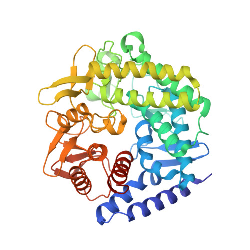
Computational Design of Experiment Unveils the Conformational Reaction Coordinate of GH125 alpha-Mannosidases.
Alonso-Gil, S., Males, A., Fernandes, P.Z., Williams, S.J., Davies, G.J., Rovira, C.(2017) J Am Chem Soc 139: 1085-1088
- PubMed: 28026180
- DOI: https://doi.org/10.1021/jacs.6b11247
- Primary Citation of Related Structures:
5M7I, 5M7Y - PubMed Abstract:
Conformational analysis of enzyme-catalyzed mannoside hydrolysis has revealed two predominant conformational itineraries through B 2,5 or 3 H 4 transition-state (TS) conformations. A prominent unassigned catalytic itinerary is that of exo-1,6-α-mannosidases belonging to CAZy family 125. A published complex of Clostridium perfringens GH125 enzyme with a nonhydrolyzable 1,6-α-thiomannoside substrate mimic bound across the active site revealed an undistorted 4 C 1 conformation and provided no insight into the catalytic pathway of this enzyme. We show through a purely computational approach (QM/MM metadynamics) that sulfur-for-oxygen substitution in the glycosidic linkage fundamentally alters the energetically accessible conformational space of a thiomannoside when bound within the GH125 active site. Modeling of the conformational free energy landscape (FEL) of a thioglycoside strongly favors a mechanistically uninformative 4 C 1 conformation within the GH125 enzyme active site, but the FEL of corresponding O-glycoside substrate reveals a preference for a Michaelis complex in an O S 2 conformation (consistent with catalysis through a B 2,5 TS). This prediction was tested experimentally by determination of the 3D X-ray structure of the pseudo-Michaelis complex of an inactive (D220N) variant of C. perfringens GH125 enzyme in complex with 1,6-α-mannobiose. This complex revealed unambiguous distortion of the -1 subsite mannoside to an O S 2 conformation, matching that predicted by theory and supporting an O S 2 → B 2,5 → 1 S 5 conformational itinerary for GH125 α-mannosidases. This work highlights the power of the QM/MM approach and identified shortcomings in the use of nonhydrolyzable substrate analogues for conformational analysis of enzyme-bound species.
Organizational Affiliation:
Departament de Química Inorgànica i Orgànica (Secció de Química Orgànica) & Institut de Química Teòrica i Computacional (IQTCUB), Universitat de Barcelona , 08028 Barcelona, Spain.


