5M1M

Crystal structure of matrix protein 1 from Influenza C virus (strain C/Ann Arbor/1/1950)


Experimental Data Snapshot

  • Method: X-RAY DIFFRACTION
  • Resolution: 1.50 Å
  • R-Value Free: 0.220 
  • R-Value Work: 0.188 
  • R-Value Observed: 0.189 

wwPDB Validation   3D Report Full Report


This is version 1.1 of the entry. See complete history


Literature

The Matrix protein M1 from influenza C virus induces tubular membrane invaginations in an in vitro cell membrane model.

Saletti, D.Radzimanowski, J.Effantin, G.Midtvedt, D.Mangenot, S.Weissenhorn, W.Bassereau, P.Bally, M.

(2017) Sci Rep 7: 40801-40801

  • DOI: https://doi.org/10.1038/srep40801
  • Primary Citation of Related Structures:  
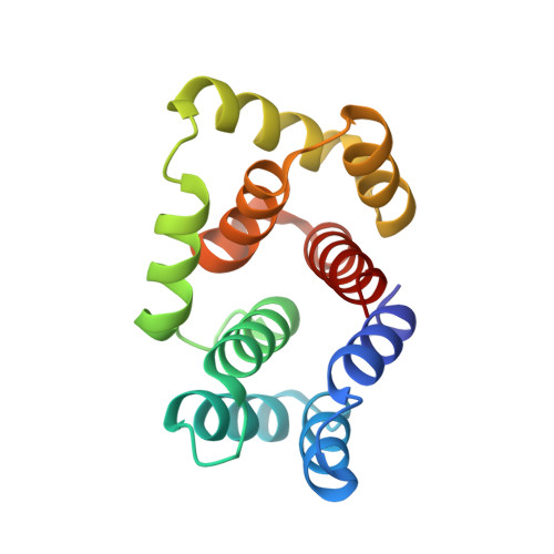
    5M1M

  • PubMed Abstract: 

    Matrix proteins from enveloped viruses play an important role in budding and stabilizing virus particles. In order to assess the role of the matrix protein M1 from influenza C virus (M1-C) in plasma membrane deformation, we have combined structural and in vitro reconstitution experiments with model membranes. We present the crystal structure of the N-terminal domain of M1-C and show by Small Angle X-Ray Scattering analysis that full-length M1-C folds into an elongated structure that associates laterally into ring-like or filamentous polymers. Using negatively charged giant unilamellar vesicles (GUVs), we demonstrate that M1-C full-length binds to and induces inward budding of membrane tubules with diameters that resemble the diameter of viruses. Membrane tubule formation requires the C-terminal domain of M1-C, corroborating its essential role for M1-C polymerization. Our results indicate that M1-C assembly on membranes constitutes the driving force for budding and suggest that M1-C plays a key role in facilitating viral egress.


  • Organizational Affiliation

    Laboratoire Physico Chimie Curie, Institut Curie, PSL Research University, CNRS UMR168, 75005, Paris, France.


Macromolecules
Find similar proteins by:  (by identity cutoff)  |  3D Structure
Entity ID: 1
MoleculeChains Sequence LengthOrganismDetailsImage
Matrix protein 1155Influenza C virus (C/Ann Arbor/1/50)Mutation(s): 0 
UniProt
Find proteins for Q6I7B9 (Influenza C virus (strain C/Ann Arbor/1/1950))
Explore Q6I7B9 
Go to UniProtKB:  Q6I7B9
Entity Groups  
Sequence Clusters30% Identity50% Identity70% Identity90% Identity95% Identity100% Identity
UniProt GroupQ6I7B9
Sequence Annotations
Expand
  • Reference Sequence
Experimental Data & Validation

Experimental Data

  • Method: X-RAY DIFFRACTION
  • Resolution: 1.50 Å
  • R-Value Free: 0.220 
  • R-Value Work: 0.188 
  • R-Value Observed: 0.189 
  • Space Group: C 1 2 1
Unit Cell:
Length ( Å )Angle ( ˚ )
a = 66.398α = 90
b = 26.841β = 108.4
c = 77.224γ = 90
Software Package:
Software NamePurpose
PHENIXrefinement
XDSdata reduction
SCALAdata scaling
PHENIXphasing

Structure Validation

View Full Validation Report


Entry History & Funding Information

Deposition Data


Funding OrganizationLocationGrant Number
French National Research AgencyFrance--

Revision History  (Full details and data files)

  • Version 1.0: 2017-02-08
    Type: Initial release
  • Version 1.1: 2017-09-06
    Changes: Author supporting evidence, Data collection