5L46

Crystal structure of human dimethylglycine-dehydrogenase


Experimental Data Snapshot

  • Method: X-RAY DIFFRACTION
  • Resolution: 3.09 Å
  • R-Value Free: 0.269 
  • R-Value Work: 0.179 
  • R-Value Observed: 0.183 

wwPDB Validation   3D Report Full Report


Ligand Structure Quality Assessment 


This is version 1.3 of the entry. See complete history


Literature

Structure and biochemical properties of recombinant human dimethylglycine dehydrogenase and comparison to the disease-related H109R variant.

Augustin, P.Hromic, A.Pavkov-Keller, T.Gruber, K.Macheroux, P.

(2016) FEBS J 283: 3587-3603

  • DOI: https://doi.org/10.1111/febs.13828
  • Primary Citation of Related Structures:  
    5L46

  • PubMed Abstract: 

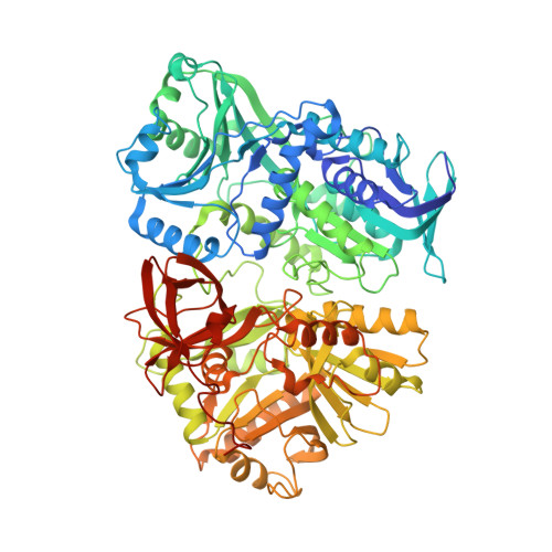
    The human dimethylglycine dehydrogenase (hDMGDH) is a flavin adenine dinucleotide (FAD)- and tetrahydrofolate (THF)-dependent, mitochondrial matrix enzyme taking part in choline degradation, one-carbon metabolism and electron transfer to the respiratory chain. The rare natural variant H109R causes dimethylglycine dehydrogenase deficiency leading to increased blood and urinary dimethylglycine concentrations. A detailed biochemical and structural characterization of hDMGDH was thus far hampered by insufficient heterologous expression of the protein. In the present study, we report the development of an intracellular, heterologous expression system in Komagataella phaffii (formerly known as Pichia pastoris) providing the opportunity to determine kinetic parameters, spectroscopic properties, thermostability, and the redox potential of hDMGDH. Moreover, we have successfully crystallized the wild-type enzyme and determined the structure to 3.1-Å resolution. The structure-based analysis of our biochemical data provided new insights into the kinetic properties of the enzyme in particular with respect to oxygen reactivity. A comparative study with the H109R variant demonstrated that the variant suffers from decreased protein stability, cofactor saturation, and substrate affinity.


  • Organizational Affiliation

    Institute of Biochemistry, Graz University of Technology, Austria.


Macromolecules
Find similar proteins by:  (by identity cutoff)  |  3D Structure
Entity ID: 1
MoleculeChains Sequence LengthOrganismDetailsImage
Dimethylglycine dehydrogenase, mitochondrial
A, B
841Homo sapiensMutation(s): 0 
Gene Names: DMGDH
EC: 1.5.8.4
UniProt & NIH Common Fund Data Resources
Find proteins for Q9UI17 (Homo sapiens)
Explore Q9UI17 
Go to UniProtKB:  Q9UI17
PHAROS:  Q9UI17
GTEx:  ENSG00000132837 
Entity Groups  
Sequence Clusters30% Identity50% Identity70% Identity90% Identity95% Identity100% Identity
UniProt GroupQ9UI17
Sequence Annotations
Expand
  • Reference Sequence
Small Molecules
Ligands 1 Unique
IDChains Name / Formula / InChI Key2D Diagram3D Interactions
FAD
Query on FAD

Download Ideal Coordinates CCD File 
C [auth A],
D [auth B]
FLAVIN-ADENINE DINUCLEOTIDE
C27 H33 N9 O15 P2
VWWQXMAJTJZDQX-UYBVJOGSSA-N
Experimental Data & Validation

Experimental Data

  • Method: X-RAY DIFFRACTION
  • Resolution: 3.09 Å
  • R-Value Free: 0.269 
  • R-Value Work: 0.179 
  • R-Value Observed: 0.183 
  • Space Group: P 1 21 1
Unit Cell:
Length ( Å )Angle ( ˚ )
a = 83.377α = 90
b = 119.868β = 92.58
c = 86.466γ = 90
Software Package:
Software NamePurpose
PHENIXrefinement
XDSdata reduction
XDSdata scaling
PHASERphasing

Structure Validation

View Full Validation Report


Ligand Structure Quality Assessment 


Entry History & Funding Information

Deposition Data


Funding OrganizationLocationGrant Number
Austrian Science FundAustriaW901

Revision History  (Full details and data files)

  • Version 1.0: 2016-08-17
    Type: Initial release
  • Version 1.1: 2016-10-19
    Changes: Database references
  • Version 1.2: 2017-09-06
    Changes: Author supporting evidence
  • Version 1.3: 2024-01-10
    Changes: Data collection, Database references, Refinement description